Download
1 / 24
240 likes | 597 Vues
PFOS-015 CELLULAR ORGANELLES. 1. Introduction 2. Nucleus 3. Endoplasmic reticulum 4. Golgi apparatus 5. Lysosomes 6. Mitochondria 7. Peroxisomes 8. Final Remarks. Prof. K.M. Chan Rm 513B, Basic Medical Science Building Department of Biochemistry Chinese University Tel: 3163-4420

E N D
PFOS-015 CELLULAR ORGANELLES 1. Introduction 2. Nucleus 3. Endoplasmic reticulum 4. Golgi apparatus 5. Lysosomes 6. Mitochondria 7. Peroxisomes 8. Final Remarks Prof. K.M. Chan Rm 513B, Basic Medical Science Building Department of Biochemistry Chinese University Tel: 3163-4420 Email: kingchan@cuhk.edu.hk
1. Introduction • Viruses and phage- show no cell membrane, their genetic materials either DNA or RNA are wrapped within the protein coat. They rely on cells to replicate. • Cells: prokaryotic cells, eukaryotic cells and archeons (archae bacteria) • Biomolecules/chemicals wrapped inside membrane: primitive cell • Archaeon (Archaebacteria) found have ether-lipids with branched chains to live in high temperature vent by volcano outlet. • Did life emerged from lava rock during volcano outbreak in the ocean to wrap up some useful “chemical soap” by chance?
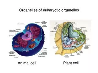
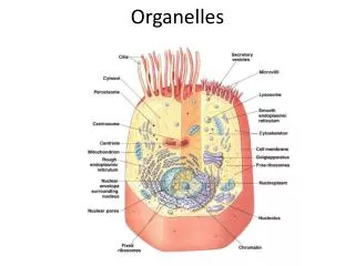
Membranes wrap the cells and their organelles inside • Membrane is a selectively permeablebarrier between the cell and the external environment. • The lipid bilayer structure defines inner membrane and outer membrane sides. Two sides of the membrane show different properties. • Homeostasis- selective permeability allows the cell to maintain a constant internal environment. • For eukaryotic cells, membranes also define cellular organelles to carry out different specific functions at a well defined location: compartmentation.
1.2 The E. coli. Cell and Human Cell • A primitive cell like bacterium E .coli cell has a single compartment in which DNA, proteins, lipids and the biomolecules are crowded together in a well organized way. • The cell is small, simple, it grows fast and can adapt to rapid environmental changes. • In contrast, eukaryotic cells like human cells are a millions times larger, much more complex, and contain organelles to support different cellular functions from digestion to generation of energy and storage of nutrients. • Human cells develop to specific cell types in different organs (differentiations). Specific cell types for specific functions.
Size Range of Cells • Small molecules (chemicals) < 1 nm (10-9 m) • Proteins and lipids- biomolecules < 10 nm • Ribosomes= 20 nm; viruses < 100 nm • Bacteria nm, 1 µm to 10 µm (10-6 m) • Mitochondrion, 2 µm; Nucleus, 7 µm • Plant and animal cells: 10-100 µm • Frog egg, 2 mm; salmon egg, 6 mm • Chicken egg, 4-5 cm • Length of muscle and nerve cells: 50-70 cm.
Bacterial cells have thick cell wall made of carbohydrates, could be differentiated as Gram + and – cells by Gram’s stain. Mesosome is infolding of membrane for specialized functions Cell wall Capsule http://www.bact.wisc.edu/themicrobialworld/structure.html Mesosome Membrane Nucleoid (DNA) region Ribosomes Peptidoglycan sugars plus amino acids Contains lipo- polysaccharides (LPS) which are endotoxins to animals in the outer membrane Flagella
May differentiate to different cell-types Adapted from the Merck Manuals On-line Medical Library: http://www.merck.com/mmhe/sec01/ch001/ch001b.html#sec01-ch001-ch001b-6
2. The Nucleus • Double membrane envelope: outer membrane continues with endoplasmic reticulum. • Nuclear pores to communicate with outside of the nucleus. “Gates” are necessary to ensure the various events impinging on gene transcription and cell signaling. • Luminal subunits in between the outer and inner membrane with a ring structure and nuclear cage. • Nucleolus contains chromatin actively transcribe ribosomal RNAs (rRNAs).
Nuclear translocator protein (vehicle) Architectures of nucleus Pore complex Outer membrane Inner membrane Luminal subunit Chromatin:DNA with histones and non-histone proteins Nuclear pores Ribosome: RNA translated to protein Inner membrane Outer Nuclear membrane Rough Endoplasmic Reticulum Nucleolus: ribosomal RNA genes are being transcribed to make ribosomal RNAs.
3. Endoplasmic Reticulum (ER) • Spans the entire cell. • Attaches with numerous ribosomes for protein translation (Rough ER). • The proteins made in the RER are folded, may be glycosylated (post-translational modifications by adding sugars), and sorted to various parts of the cell. • ER also produces phospholipids (smooth ER). • As a depot of calcium ions (smooth ER) in the cell for controlling the signaling of cellular processes.
Rough ER dedicated for protein production http://cellbio.utmb.edu/cellbio/ribosome.htm#Ribosome-Endoplasmic%20Reticulum From RER to Golgi Apparatus http://cellbio.utmb.edu/cellbio/golgi.htm
4. The Golgi Apparatus • Proteins made in the RER are sorted to the Golgi apparatus for further processing. • They enter the Golgi at the cis face from vesicles formed at the ends of the ER. • In the cisternae of the Golgi, proteins are labeled (secondary modification), sorted and delivered to the trans face of the Golgi apparatus. http://sun.menloschool.org/~birchler/cells/animals/golgi/structure.html
Tunicamycin • Proteins are glycosylated in ER before dispatch to the Golgi apparatus. • It acts by mimicking the structure of UDP-N-acetylglucosamine, the substrate in the first enzymatic step in the glycosylation pathway. • It thus blocks protein post-translational modification to kill eukaryotic cells; • It’s an antibiotics.
Protein sorting (trafficking) • As vesicles formed in the Golgi, then sent to proper locations for specific function to be carried out in other parts of the cells (e.g. organelles or stored as secretory vesicles). • Proteins in vesicles would accumulate and wait for signal to be exported out by exocytosis (either regulated or constitutively secreted out of the cells. • Some proteins are delivered to and kept on cell membrane.
Rough Endoplasmic Reticulum and Golgi Apparatus Lysosome Early endosome Other organelles Rough Endoplasmic Reticulum Engulfed materials Regulated secretary pathways Vesicle Trans Golgi apparatus ECOSYTOSIS Cis-Golgi apparatus Constitutive secretary pathway Golgi Apparatus Proteins are further modified and sorted in the Golgi apparatus
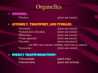
5. The Lysosome • A vesicle with acidic condition to digest and remove unwanted materials, or break down materials for cellular uptake or re-use. • It contains digestive enzymes (primary lysosome). • After bud off from the Golgi, the primary lysosomes fuse with autophagic vesicles to form phagocytic vacuoles (secondary lysosomes), or engulfed materials to form endosomes. • Autophagy: degradation of intracellular components in lysosomes; heterophagy: degradation of phagocytic materials (in-coming from outside). • The debris could be discarded outside of the cell or kept as granules or recycle to cytoplasm.
LYSOSOMAL HYDROLASES • Synthesized in the ER, lysosomal hydrolases were moved to the cis Golgi network where they are covalently modified by the addition of Mannos-6-Phosphate (M6P) • M6P receptor recognizes the labelled hydrolases in trans-Golgi and move them to the endosomes • I-Cell disease is a genetic defect that produce the enzymes without M6P. Hence the enzymes could not be deposited to the endosomes for digestions. • Glycosaminoglycans and glyco-lipids accumulated in the lysosomes (without proper digestions) and the patients exhibit psychomotor retardation and skeletal deformities. They die early, usually before age 8.
Tay-Sachs Disease • It’s a lysosome storage disease • lacks of hexosaminidase A. • Results in the abnormal accumulation of other cellular materials in the brain. • Mental retardation and blindness resulted from Ganglioside GM2 accumulation • Also known as GM2 gangaliosidosis or Sphingolipidosis • The lysosomes cannot function properly or even break to release not just digestive enzymes, but also acid in the cells to create a very acidic condition killing the neural cells.
Gout and Rheumatoid Arthritis • Gout is deposit of uric acid crystals of the joints often from over consumption of meat; and Rheumatoid factor complexes in the leucocytes of the joints. • Break lysosome to release enzymes that degrade the components of the synovial membrane, resulting in great pain and joint deformation.
6. The MITOCHONDRION • Double membrane of inner and outer. • Inner membrane fold to create the matrix space and inter-membrane space in between inner and outer membrane. • The matrix contains enzymes to oxidize metabolites. • The inter-membrane has potential gradient generated by pumps at the inner membrane, energy released from the gradient produce ATP energy.
Outer membrane, with channel forming proteins but only permeable to 5 kDa molecules or less. Other enzymes on this membrane facilitate metabolism of lipid for use in matrix and transport of specially required protein in the mitochondrion. MAJOR FUNCTION OF MITOCHONDRION: OXIDATIVE PHOSPHORYLATION (oxidation of NADH to produce ATP) Matrix, internal space with enzymes for oxidation of pyruvate and fatty acids and for the citric acid cycle (Kreb’s cycle). Cristae in boxed region Intermembrane space, for enzymes using ATP passing out of the matrix to phosphorylate other nucleotides. Inner membrane, folded into numerous cristae to increase total surface area for electron-transport chain (respiratory enzymes), ATP synthase and transporters for metabolites going through the matrix.
Mitochondrial (mt) disease • Mt has its own DNA to make its own proteins; with some imported proteins from cytoplasm • Incorporated into eukaryotic cells by endosymbiotic processes during the course of evolution. • Mt disease can arise from defective import of proteins (e.g. heat shock protein 70 needed for docking and folding of proteins in the mt) or mutations in mt genes. • Tissues affected are those heavily dependent on ATP, e.g. nerve and muscle cells; resulted in mental retardation and muscular weakness. • For those need many mt (mt-gene) for function, Leber’s hereditary optic neuropathy and Kearns-Sayre Syndrome are caused by mutation of NADH-CoQ reductase gene; progressive blindness, abnormal heart beat and nerve degeneration resulted.
7. Peroxisomes • An organelle for oxidation of fatty acids and toxicants • Abundant in hepatocytes (liver cells), where oxidation of fatty acids (and other organic matters) takes place and produces hydrogen peroxide; • Mainly produce catalase to decompose hydrogen peroxide into water. • Zellweger syndrome is caused by the mistakes of protein import into the peroxisomes and hence accumulation of long chain fatty acids and pristanic acids in plasma and tissues. • The defects can lead to severe impairment of many organs and death.
8. Final Remarks: • Eukaryotic cells are highly organized with membranous organelle-as isolated compartments to carry out specific life processes and biological functions. • The nucleus contains DNA for gene transcription and production of RNAs. Transcriptional factors and signals go in and out through nuclear pores under a tight control. • Rough ER handles translation and post-translational modification, protein sorting, vesicle formation are done in Golgi apparatus. • Mitochondiron is responsible for respiration and ATP energy production; it has 2 membranes and its own genes. • Lysosomes are for cellular digestions, peroxisomes are for fatty acid oxidation and detoxification or Redox reactions of intoxicated chemicals.