Download
1 / 1
10 likes | 140 Vues
Serosurveillance for Anaplasma phagocytophilum Antibodies in White-tailed Deer in Iowa. Acknowledgements. Results. Discussion. Materials & Methods. Introduction.

E N D
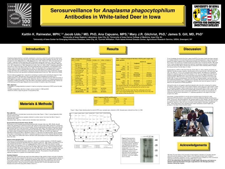
Serosurveillance for Anaplasma phagocytophilum Antibodies in White-tailed Deer in Iowa Acknowledgements Results Discussion Materials & Methods Introduction Kaitlin K. Rainwater, MPH,1,4 Jacob IJdo,2 MD, PhD, Ana Capuano, MPS,3 Mary J.R. Gilchrist, PhD,1 James S. Gill, MD, PhD1 1University of Iowa Hygienic Laboratory, Iowa City, IA; 2University of Iowa Carver College of Medicine, Iowa City, IA; 3University of Iowa Center for Emerging Infectious Diseases, Iowa City, IA; 4Current Affiliation: Plum Island Animal Disease Center, Agricultural Research Service, USDA, Greenport, NY • Anaplasma phagocytophilum is primarily maintained in nature by a rodent-tick cycle with the white-footed mouse (Peromyscus leucopus) serving as the rodent reservoirand Ixodes scapularis, the deer tick, as the principal tick vector (Magnarelli et al. 1995, Magnarelli et al. 1997, Telford et al. 1996).The preferred host of adult I. scapularis is white-tailed deer (WTD [Odocoileus virginianus]) (Lane et al. 1991), which have been shown to serve as competent sentinel animals for the surveillance of A. phagocytophilum (Belongia et al. 1997, Magnarelli et al. 1999, Magnarelli et al. 2004, Tate et al. 2005). While I. scapularis populations are known to exist in the state of Iowa, they have historically been concentrated in the northeastern and eastern areas of the state (Bartholomew et al. 1995). The assumption that A. phagocytophilum is present in these regions of Iowa is consistent with the fact that the state shares its northeastern and northern borders with Wisconsin and Minnesota, respectively, where the initial human cases of HGA were reported in 1994 and continue to be reported at consistently elevated levels (Chen et al. 1994, Demma et al. 2005). • Recent evidence suggests that I. scapularis is increasing in numbers and is extending its range into Iowa (Lingren et al. 2005). Although there are no studies specifically investigating the presence of A. phagocytophilum in Iowa, five cases of HGA have been reported in Iowa since the year 2000 (University of Iowa Hygienic Laboratory, unpublished data). While this may be an indication of increased awareness and recognition of HGA by physicians, it may also reflect a geographic expansion beyond the two endemic foci of the Northeast and the upper Midwest and a true increase in incidence rates of the disease. • Main objectives: • Investigate if A. phagocytophilum is present in Iowa by conducting a serosurvey of WTD across the state in 2004 • Compare seropositivity rates from a single, common location in 1999 and 2004 • Assess the effects of age and gender on seropositivity rates To our knowledge this study is the first to report that WTD in the state of Iowa have been exposed to A. phagocytophilum, thus expanding the previously identified geographic range of this organism beyond the endemic adjacent area of Minnesota and Wisconsin (Demma et al. 2005). Eleven of the 13 sites in this serosurvey revealed at least one positive serum sample by ELISA or WB. Eight of the 13 sites showed positive sera by both methods. Interestingly, comparison of seropositivity rates at F.W. Kent Park (Site 3) and the Iowa City area (Site 4) were drastically different despite the close proximity of the two sites (13 miles). Seroprevalence of WTD in F.W. Kent Park was 76.9% (95% CI = 54% to 99.8%) by ELISA, the highest of any site surveyed. However, seroprevalence at the Iowa City site in the same year was only 1.5% (95% CI = 0% to 3.7%) using the same testing method. This difference is significant despite the relatively small F.W. Kent Park sample (n=13). This may suggest that some sites are relatively isolated and differences in the surrounding environment may affect transmission of A. phagocytophilum. While the area in which Iowa City samples were collected is more urban with heavy traffic, housing, and businesses, F.W. Kent Park is a densely wooded, rural area that may have significant populations of I. scapularis and P. leucopus. We intend to perform future studies in this area that will include dragging and flagging for ticks and small mammal capture in order to determine the significance of these populations. Although the data do not suggest a significant adjusted odds ratio when comparing the eastern and western regions of Iowa, part of the variation between regions was due to age variation (Table 3). The analysis indicates a higher seroprevalence rate among adult WTD in Iowa. In fact, among eastern Iowa adult WTD, we observed increased odds compared to adults of other regions. We expect the seroprevalence rate in central Iowa adult WTD to rise over the next several years in conjunction with the recent identification of I. scapularis populations in this region of the state (Lingren et al. 2005). Importantly, A. phagocytophilum is now the second ehrlichial agent to be found in Iowa. Previously, WTD were found to have antibodies to Ehrlichia chaffeensis (Mueller-Anneling et al. 2000). Furthermore, Borrelia burgdorferi, the causative agent of Lyme disease, has been identified in I. scapularis ticks in Iowa (Lingren et al. 2005) and several laboratory-confirmed cases of Lyme disease are reported in Iowa each year (University of Iowa Hygienic Laboratory, unpublished data). These observations have important public health implications of which health care providers in the state of Iowa should be aware. Figure 1. Map of Iowa indicating sites from which WTD serum samples were collected in 2004. Samples were collected from Site 4 in 1999. • Sera collection • 628 WTD at thirteen controlled deer harvest sites across Iowa (Figure 1, Table 1) during September 2004 through February 2005. • 282 previously untested serum samples collected in a similar manner from Iowa City (Site 4, Figure 1) during the fall of 1999. • Age group (fawn, yearling, or adult) and sex information were determined. • Enzyme-linked ImmunoSorbent Assay (ELISA) • Performed on all 910 samples as previously described with changes (IJdo et al. 1999). Briefly, the p44 antigen was generated as a p44 fusion protein with maltose binding protein (MBP) produced in Escherichia coli and purified by affinity column chromatography. Ninety-six-well polystyrene plates were coated with p44-MBP, blocked with 5% nonfat milk solution, and washed with PBS-Tween. Sera diluted in PBS were added to duplicate wells, plates incubated and washed as described above. HRP-conjugated rabbit anti-deer IgG was added, plates incubated and washed as described above. TMB and 1N H2SO4 was added and absorbance measured at 405nm. Absorbance values of 0.300 were recorded as positive. Serum samples at four sites in southwestern Iowa where I. scapularis populations have not been reported (Bartholomew et al. 1995, Lingren et al. 2005) were used to calculate cutoff values based on three standard deviations (SD) above the mean. • Western Immunoblotting (WB) • Performed on all ELISA-positive serum samples (n=62) and on a random selection of 36 ELISA-negative serum samples as previously described with minor variations (IJdo et al. 1999). Briefly, cell lysates of HL-60 cells infected with the NCH-1 strain of A. phagocytophilum (Telford et al. 1996) were placed in sample buffer, heated, and separated on a 12% SDS-PAGE gel. Proteins transferred to nitrocellulose and unbound sites were blocked using 3% bovine serum albumin (BSA). Blots were cut into strips and probed with deer sera diluted in PBS. Strips were washed three times with PBS-Tween, placed in HRP-conjugated rabbit anti-deer immunoglobulin G diluted in PBS-Tween, washed as described above, placed in SuperSignal West Femto Maximum Sensitivity Substrate, and exposed to Xray film. The presence of the p44 band was required as evidence of a positive immunoblot result (Figure 2). • Statistical Analysis • A two-sided Fisher’s exact test was used in the initial analysis of age, gender, location and year of collection. Binomial intervals were computed for risk factors for the crude prevalence of A. phagocytophilum infection in WTD. For data collected in 2004, unadjusted and adjusted odds ratios were computed for each risk factor using logistic regression. A final multivariable model was defined using a saturated model and manual backward elimination. Ninety-five percent confidence intervals (95% CI) were calculated at each step. Analyses were performed using SAS software version 9.1. Figure 2. Representative immunoblots of deer sera collected in Iowa showing reactivities to the p44 antigen of A. phagocytophilum. Molecular mass standards are shown in kilodaltons along the left. Lanes marked with (-) or (+) are negative and positive controls, respectively. Lanes 2 and 3 show WTD serum samples with antibodies to the p44 antigen, and lanes 1 and 4 are negative serum samples. We thank Thomas Gahan of the University of Iowa Hygienic Laboratory for providing relevant data and technical assistance; Adam Carlson of the University of Iowa Carver College of Medicine for laboratory assistance; and Dr. Louis Magnarelli and Tia Mastrone of the Connecticut Agricultural Experiment Station for providing WTD serum controls. K.K.R. was supported by the Association of Public Health Laboratories and Centers for Disease Control and Prevention through an appointment to the Emerging Infectious Diseases Fellowship Program.