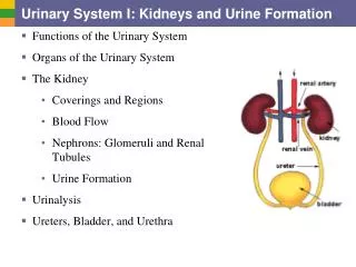
Urine Formation PPT
0 likes | 266 Vues
Urine formation is an intricate and vital process that takes place in our kidneys. It involves the filtration of blood, reabsorption of essential substances, and the secretion of waste products. This remarkable mechanism ensures the balance of fluids and electrolytes in our bodies, aiding in the maintenance of overall health.

Urine Formation PPT
E N D
Presentation Transcript
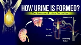
Urine formation involves three main processes ⓵ ⓵Glomerular filtration Glomerular filtration ⓶ ⓶ Reabsorption Reabsorption ⓷ ⓷Tubular Secretion
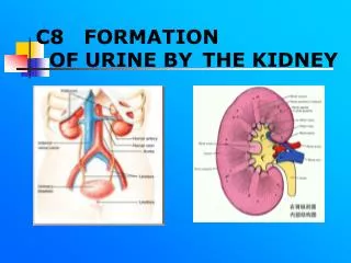
⓵ ⓵GLOMERULAR FILTRATION GLOMERULAR FILTRATION ➢ The first step in urine formation is the filtration of blood, which is carried out by the glomerulus ➢ This process occurs in the Malpighian corpuscle of the nephron ➢ The filtration membrane consist of 3 layers: 1. Endothelium of glomerular blood vessels. 2. Epithelium of Bowman’s capsule. 3. Basement membrane between these two layers. ➢ Epithelial cells (podocytes) of the Bowman’s capsule are arranged in an intricate manner leaving some minute spaces called filtration slits (slit pores). ➢ Almost all constituents of blood plasma except the proteins pass onto lumen of Bowman’s capsule. Therefore, it is considered as a process of ultra filtration
⓵ ⓵GLOMERULAR FILTRATION GLOMERULAR FILTRATION ➢ The plasma fluid filters out from glomerular capillaries into bowman's capsule of nephrons is called glomerular filtrate ➢ About 1100-1200 ml of blood is filtered by kidneys per minute. It constitutes 1/5th of the blood pumped out by each ventricle of the heart in a minute. ➢ The amount of glomerular filtrate formed per minute is called Glomerular filtration rate (GFR). ➢ Normal GFR = 125 ml/minute, i.e., 180 litres/day.
⓶ ⓶ REABSORPTION REABSORPTION ➢ 180 litres of glomerular filtrate is produced daily. But 99% of this is reabsorbed by renal tubules. So normal volume of urine released is 1.5 litres. ➢ From the filtrate, glucose, amino acids, Na+, etc. are reabsorbed actively and nitrogenous wastes are absorbed passively. ➢ Passive reabsorption of water occurs in the initial segments of the nephron. ➢ PCT reabsorbs most of the nutrients, and 70-80% of electrolytes & water.
⓶ ⓶ REABSORPTION REABSORPTION ➢ Simple cuboidal brush border epithelium of PCT increases surface area for reabsorption. ➢ Loop of Henle maintains high osmolarity of medullary interstitial fluid. ➢ Descending limb is permeable to water but almost impermeable to electrolytes. This concentrates the filtrate. ➢ In Ascending limb, minimum reabsorption occurs. It is impermeable to water but allows transport of electrolytes. So, filtrate gets diluted.
⓶ ⓶ REABSORPTION REABSORPTION ➢ In DCT, conditional reabsorption of Na+ & water takes place. ➢ Collecting duct extends from cortex to inner parts of medulla. It reabsorbs large amount of water to concentrate urine. ➢ It also allows passage of small amounts of urea into medullary interstitium to keep up the osmolarity.
⓷ ⓷TUBULAR SECRETION ➢ Cells of PCT & DCT maintain ionic (Na & K balance) and acid-base balance (pH) of body fluids by selective secretion of H+, K+ & NH3 into the filtrate and absorption of HCO3- from it. ➢ Collecting duct maintains pH & ionic balance of blood by the secretion of H+ and K+ ions. ➢ Some drugs are filtered in the glomerulus and so are actively secreted into the filtrate during the tubular secretion
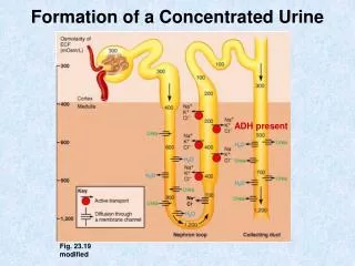
➢ Mammals have the ability to produce a concentrated urine. ➢ The Henle's loop and vasa recta play a significant role in this. ➢ There are two counter current mechanisms inside the kidneys. I. The flow of filtrate in the two limbs of Henle's loop is in opposite directions and thus forms a counter current. II. The flow of blood through the two limbs of vasa recta is also in a counter current pattern ➢ Due to the counter current and proximity between Henle’s loop & vasa recta, osmolarity increases from cortex (300 mOsmolL-1) to the inner medullary interstitium (1200 mOsmolL-1). This gradient is caused by NaCl & urea.
➢ The descending limb of the loop of Henle is permeable to water but not salts and ascending limb of the loop of Henle is permeable to salts but not water. ➢ NaCl is transported by ascending limb of Henle’s loop that is exchanged with descending limb of vasa recta. NaCl is returned to interstitium by ascending limb of vasa recta. ➢ Similarly, small amount of urea enter the thin segment of the ascending limb of Henle’s loop which is transported back to the interstitium by the collecting tubule.
➢ Thus electrolytes and urea are retained in the interstitium and maintain a concentration gradient (interstitial gradient) in medullary interstitium. ➢ The loop of Henle establishes a salt gradient (hypertonicity) in the medulla ➢ It enables easy passage of water from collecting tubule to concentrate the filtrate (urine). ➢ Thus DCT & collecting duct produce urine four times concentrated than the initial filtrate formed.
➭CLICK HERE FOR NEXT PART ➭CLICK HERE FOR VIDEO LECTURES ➭CLICK HERE FOR OTHER CHAPTERS