SlideServe
Menu
Préférences
© SlideServe · v2026

Unraveling Ribosome Structure: The Pioneering Work of Prof. Ada Yonath

ralph avatar
21 diapositives · 345 Views · Sep 27, 2014

Scientists often face challenges in visualizing structures smaller than the wavelength of visible light, such as ribosomes. Prof. Ada Yonath made significant strides in this area using X-ray crystallography to investigate ribosome organization and protein composition. Her innovative approach involved crystallizing bacterial ribosomes and maintaining stability during X-ray exposure. This work not only led to her Nobel Prize in Chemistry in 2009 but also enhanced our understanding of how antibiotics target bacterial ribosomes, paving the way for more effective drug development.
Unraveling Ribosome Structure: The Pioneering Work of Prof. Ada Yonath - slide 1
1 / 21

Télécharger la présentation

Unraveling Ribosome Structure: The Pioneering Work of Prof. Ada Yonath
Une image / un lien ci-dessous est fourni (tel quel) pour télécharger la présentation

Politique de téléchargement : le contenu du site vous est fourni TEL QUEL pour votre information et votre usage personnel ; il ne peut être vendu, concédé sous licence ni partagé sur d’autres sites sans l’accord de son auteur.Le contenu vous est fourni TEL QUEL, pour votre information et votre usage personnel uniquement. Téléchargez la présentation en cliquant sur ce lien. Lors du téléchargement, si vous ne parvenez pas à télécharger une présentation, l’éditeur a peut-être supprimé le fichier de son serveur.Pendant le téléchargement, si vous ne parvenez pas à obtenir une présentation, le fichier a peut-être été supprimé par l’éditeur.

F I N

Plus de contenu associé

Transcription de la présentation

  1. Scientists like to actually see the things they research. But this isn't always possible. Source:Clipart ETC

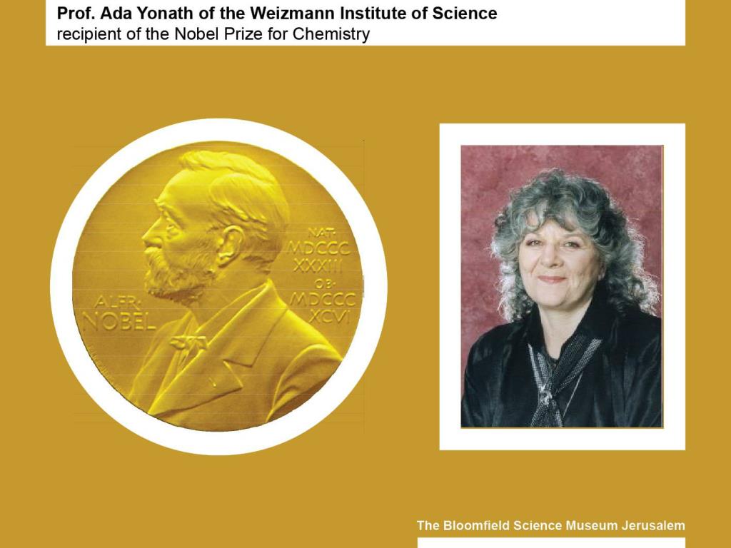
  2. Most of what we see is larger than the wavelength of visible light. Scientists occasionally want to study things that are smaller thanthe wavelength of visible light. Wavelength of ultraviolet light: 400 nanometers One nanometer = one millionth of a millimeter Source:Wikipedia

  3. That was the case with the ribosome that Nobel Prize laureate Prof. Ada Yonath wanted to explore. The diameter of a ribosome is about 20 nanometers, which is approximately 20 times smaller than the ultraviolet wavelength. Wavelength of ultraviolet light: 400 nanometers One nanometer = one millionth of a millimeter

  4. To "see" the internal structure of bodies the size of a ribosome, scientists often expose them to x-rays. This reveals the location of single atoms. The wavelength of x-rays is about 20 times smaller than the diameter of ribosomes. Source: NASA

  5. The ribosome is a key component of living cells. Source: Wikipedia

  6. Living cells have a single nucleus and thousands of ribosomes. Source:Wikipedia

  7. Ribosomes translate the genetic information within the cell's nucleus into a process for producing proteins. SourceWikipedia

  8. Proteins are huge molecules, structured like folding chains and composed of a sequence of amino acids. Source: Wikipedia

  9. Proteins are one of the most important compounds that make up living organisms. They are found in every living cell. Ribosomes themselves are composed, in part, of proteins Source:Clipart ETC

  10. Prof. Ada Yonath and other researchers wanted to know which proteins are contained in the ribosome and how they are organized spatially. To find out, they needed to rely on x-rays. Source: Prof' Ada Yonaths' Lab

  11. The method for determining the structure of biological molecules using radiation is called x-ray crystallography. Prof. Ada Yonath uses x-ray crystallography to study ribosomes. SourceNASA

  12. To prepare a living substance for x-ray crystallography research, the substance itself needs to be crystallized. Source: Prof' Ada Yonaths' Lab

  13. Different crystals diffract the x-rays directed at them in different ways. The particular composition and spatial structure of each crystal create a unique picture. This picture resembles a collection of dots. Source: Wikipedia

  14. All the pictures created during x-raying are entered into powerful computers, which feed them into a complex decoding process resulting in three-dimensional images. The computer-assisted decoding relies on mathematical formulas and hypothetical structural models, based on data gathered through other methods. Source: Wikipdia

  15. As the x-raying proceeds, it is crucial to maintain the stability of the substance being examined. In many cases, the substance is deformed due to the strong radiation it receives, similar to the way living tissues are harmed in an X-ray test. Because of the radiation, the substance changes form, preventing the researcher from determining its structure.

  16. Prof. Ada Yonath was the first to deal successfully with two complex problems: crystallizing the ribosome and preparing the crystal for x-ray without changing its form during the examination process. Source: L'OREAL-UNESCO Awards For Women in Science

  17. The living substance that Prof. Yonath used to examine ribosome structure was bacteria that live in hot springs or in the Dead Sea, which are resistant to high temperatures. Prof. Yonath cooled samples of the substance to a temperature of -185º C, preventing its decomposition under experimental conditions.

  18. This accomplishment earned her the 2009 Nobel Prize for Chemistry. She shared the prize with Thomas Steitz of Yale University in the U.S. and Venkatraman Ramakrishnan of Cambridge University in Britain. The three scientists were awarded the prize for their contribution to the study of ribosome structure and functioning.

  19. Most antibiotic medicines attach themselves to bacterial ribosomes, preventing them from acting. By understanding how antibiotics affect ribosomes, scientists can conduct research leading to the development of more effective drugs. Source: The lab of Prof' Ada Yonath

  20. Writing: Hanan Cohen Editing: Nurit Snir Graphic design: Vadik Bakman Many thanks to Prof. Ada Yonath for her comments on this presentation