Download
1 / 52
590 likes | 1.6k Vues
Eukaryotic Translation Regulation PTRM Class February 8, 2011. Differences. Translation and Transcription are uncoupled. mRNAs are monocistronic. mRNA has a 5’cap structure. mRNA has a poly(A) tail (except Histone mRNA) Ribosomes bind to the RNA at the 5’end (no Shine Dalgarno sequence).

E N D
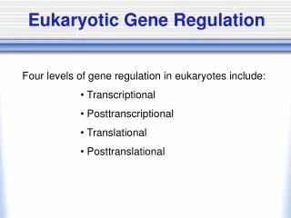
Differences Translation and Transcription are uncoupled. mRNAs are monocistronic. mRNA has a 5’cap structure. mRNA has a poly(A) tail (except Histone mRNA) Ribosomes bind to the RNA at the 5’end (no Shine Dalgarno sequence). tRNAmetiis specific for initiation in eukaryotes it is not a formylated tRNAmet Eukaryotic and Prokaryotic Translation Similarities The genetic code is nearly universal, applying to all species on our planet. Protein synthesis begins with an AUG and terminates with UGA, UAA, and UAG.
(A)n 3' mRNA Cap Poly(A) tail Secondary structures IRES Protein/RNA binding sites uORF miRNA target Localization signals AUG Kozak consensus sequence: GCC(A/G)CCAUGG Fátima Gebauer & Matthias W. Hentze Nature Reviews Molecular Cell Biology 5, 827-835 (2004)
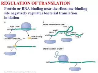
Translational Control: Mediated at the Level of Translation Initiation Prokaryotes Base pairing between the Shine Dalgarno sequence and the 3´ end of 16S rRNA facilitates translation initiation. Voet and Voet, Figure 30-42 Gebauer & Hentze Nature Reviews Molec. Cell Bio. 5, 827-835 (2004) Eukaryotes The 40S ribosomal subunit is recruited to the 5’end of the mRNA through recognition of the cap structure and interactions between translation initiation factors.
Formation of the ternary complex Formation of the 43S pre-initiation complex Recruitment of the 43S to the 5’end of the mRNA 4. 40S ribosome scans down to the start codon, positioning met-tRNAi into the P-site. 5. The 60S subunit joins, facilitated by eIF5B Scanning model of Cap-dependent Translation Initiation E P A Fátima Gebauer & Matthias W. Hentze (2004) Nature Reviews Molecular Cell Biology 5, 827-835 E P A
Role of eIF1 and eIF1A in initiation eIF1 is proposed to monitor base pairing between mRNA and Met-tRNAiMet eIF1A: maintains the fidelity of start codon recognition, likely through an interaction with eIF5 and by stabilizing the open, scanning-competent 40S conformation until the start codon is identified. Also, eIF1A may bind to the A site, blocking initiator tRNA from binding in this position. eIF1 and eIF1A enhance ternary complex loading by altering the conformation of the 40S subunit. eIF1A plays an additional role by stabilizing ternary complex once it is bound. IF3 IF1 P E A IF2 Fátima Gebauer & Matthias W. Hentze Nature Reviews Molecular Cell Biology 5, 827-835 (2004)
Model for the Functions of eIF1 and eIF1A in Eukaryotic Translation Initiation L. A. Passmore et al., Mol Cell26, 41 (Apr 13, 2007).
Closed Open n= neck b= beak sh= shoulder lf= left foot rf=right foot pt=platform • 40S-eIF1-eIF1A structure represents an open, • scanning-competent form of the initiation complex. • A new connection between the head and shoulder on the solvent side. • The ‘‘latch’’ of the mRNA entry channel is closed in empty 40S but is not visible in the 40S-eIF1-eIF1A structure. This latch is a noncovalent interaction between rRNA elements in the body Solvent surface Intersubunit surface
Clamping down of the latch following start codon recognition and eIF1 release • The density for the latch appears more pronounced in 40S-eIF1A, suggesting that the h18-h34 connection may be stronger when eIF1A is bound. Because eIF1 dissociates after start codon recognition, perhaps eIF1A, when present without eIF1, helps the 40S subunit to clamp down on the mRNA, holding it in position in preparation for 60S subunit joining. L. A. Passmore et al., Mol Cell26, 41 (Apr 13, 2007).
Recycling of the ternary complex requires eIF2B Fátima Gebauer & Matthias W. Hentze Nature Reviews Molecular Cell Biology 5, 827-835 (2004)
The ternary complex binds the 40S subunit and is recruited to the 5’end of the mRNA 3 E P A Fátima Gebauer & Matthias W. Hentze Nature Reviews Molecular Cell Biology 5, 827-835 (2004)
eIF4G is a scaffolding protein Fátima Gebauer & Matthias W. Hentze Nature Reviews Molecular Cell Biology 5, 827-835 (2004)
mRNA Circularizes Through eIF4G Binding to eIF4E and PABP http://departments.oxy.edu/biology/Stillman/bi221/091300/091300_lecture_figures.htm
eIF4E binding protein (4E-BP) Sequesters eIF4E from eIF4F complex. Apoptosis Insulin AA (Leucine) Cell Proliferation
PABP The 40S Ribosomal Subunit scans 5’ to 3’ to the initiating AUG 4A 4G 3 1A 5 1 cap 4E E P A • Start site selection • Proximity to the 5’end • Kozak consensus sequence GCC(A/G)CCAUGG • Scanning is an ATP dependent process • eIF1 and 1A are involved in scanning, eIF4A is an RNA dependent helicase; • eIF1 monitors the fidelity of codon-anticodon interaction • eIF5 promotes GTP hydrolysis of the ternary complex following recognition of the start codon. Fátima Gebauer & Matthias W. Hentze Nature Reviews Molecular Cell Biology 5, 827-835 (2004)
eIF5B Facilitates Ribosomal Subunit Joining 3 1A 5 1 E P A eIF5B E P A Fátima Gebauer & Matthias W. Hentze Nature Reviews Molecular Cell Biology 5, 827-835 (2004)
Electronmicrograph of a polysome: one mRNA strand (faint horizontal line) with many individual ribosomes attached (dark blobs). The newly synthesized polypeptide chains (proteins) can be seen as irregularly shaped extensions from the ribosomes: http://bass.bio.uci.edu/~hudel/bs99a/lecture21/polysome.gif
Polysome Analysis Freeze ribosomes onto the mRNA with cyclohexamide Load cell lysates onto a sucrose gradient. Use centrifugal force to separate mRNAs based on their translational efficiency Fractionate the gradient. Isolate total RNA from each fraction. Perform northern analysis to look at translation of specific mRNAs Polysomes Actin Northern
Toe Print Assay Pisarev et al. (2007) Methods in Enzymology 430: 147-177
What are the limits and advantages of polysome analysis and Toe Print assays?
What are the limits and advantages of polysome analysis and Toe Print assays? Toe prints don’t tell you what size the complex is: (40S or 80S) Polysomes indicate the global translation rates, where as Northern analysis can tell you the translation status of a particular mRNA Toe prints can indicate exactly where on the mRNA a complex is positioned.
Translational control by elements in the 5’UTR 1. Leaky scanning 2. IRES 2. Structure and/or Protein binding (IRE/IRP) 3. uORF (GCN4)
Leaky scanning Permits a downstream AUG to be used for initiation in preference to, or in addition to the first (5’ proximal) AUG. ORFs may be in the same or different reading frames. The primary determining feature of start codon selection is the context of the AUG: Kozak consensus sequence: GCCA/GCCAUGG Gaba et. al. (2001) EMBO J. 20:6453-6463.
Two Mechanism of Translation Initiation Cap-dependent IRES-dependent HCV FMDV E. Martínez-Salas et. al. Journal of General Virology (2001), 82, 973-984.
Cap-Independent Translation Initiation • Cellular • Cellular stress: viral infection (PKR), apoptosis, hypoxia • Normal cellular processes: M phase cell cycle (25% translation) • Viral • Genomes: Picornaviruses, Hepatitis C Virus, Cricket paralysis virus • Specific viral messages: HIV, Herpes virus (…. and the list is still growing!)
Cellular IRESes: How do they recruit the 40S subunit? • Widely assumed that the Secondary and tertiary Structure allow for interactions with translational machinery (ITAF,eIFs, 40S) • No common Structure • Composed of multiple short modules • Generally 150-300 nts long, exception: 9nt repeated element.
Dicistronic reporter Assay: A functional Assay for IRES activity Cap-dependent IRES-dependent 60S 60S 40S 40S 60S 60S IRES 5’ cap Firefly Luciferase Renilla Luciferase AAAAA 40S 40S Marla Hertz
Ribosomes enter internally Chen and Sarnow (1995) Science 268:415-417
Examples of IRES elements E. Martínez-Salas et. al. Journal of General Virology (2001), 82, 973-984.
IRESs that bind to the 40S subunit in the absence of initiation factors induce a similar conformational change in the 40S subunit as does eIF1A and eIF1 binding. Suggesting that this is a general feature of translation initiation. Opening of the exit tunnel h16 and rps3 connection mRNA entry latch 40S subunit (yellow). HCV IRES (purple) bound to a 40S subunit (yellow). C. M. Spahn et al., Science291, 1959 (Mar 9, 2001).
Protein synthesis in poliovirus-infected HeLa cells - 109 - 80 - 51 - 34 26 - 0 1 2 3 4 5 6 hrs p.i 35S-meth. pulse eIF4G-Western
Translational control by elements in the 5’UTR 1. IRES 2. Structure and/or Protein binding (IRE/IRP) 3. uORF (GCN4)
IRE/IRP: Steric Hindrance Iron is both essential and toxic so its levels must be carefully regulated within the cell. (Excess iron promotes the generation of reactive radicals, which in turn damage cells and tissues.) Transferrin receptor: Iron uptake Ferritin: required for iron storage KOSTAS PANTOPOULOS Ann. N.Y. Acad. Sci. 1012: 1–13 (2004)
eIF2 Phosphorylation Reduces Ternary Complex Starvation (GCN2) Viral infection, apoptosis (PKR) ER Stress (PERK) Haemin-regulated inhibitor (HRI) Fátima Gebauer & Matthias W. Hentze Nature Reviews Molecular Cell Biology 5, 827-835 (2004)
GCN4: Encodes a transcription factor for amino acid biosynthesis Non-starvation Starvation
A mechanism of translation initiation in which ribosomes bind to the mRNA in a cap-dependent manner (or at the 5’end) but then "jump" over large regions of the mRNA containing RNA secondary structure, upstream AUGs and open reading frames to "land" at an AUG or upstream of the initiator AUG. Shunting/Re-initiation • Shunting • Re-initiation • Shunting/re-initiation • Termination dependent re-initiation • stop-start
Shunting Examples: Adenovirus (late mRNAs) Sendaivirus (Y mRNAs) Papillomavirus (E1 mRNA) Duck hepatitis B virus mammalian mRNAs Properties: Cap (or other mechanism) RNA secondary structure Motifs in the donor region that tether the 40S to the RNA (some). Some have uORF and require termination of translation L. A. Ryabova and T. Hohn, Genes Dev14 (7), 817 (2000).
Shunting Ribosomes bind to the 5’end 40S scans down the message. Some, translate an uORF and terminate at a stop codon. 40S subunit resumes scanning The 40S subunit “shunts” to a downstream sequence. Translation initiates at a downstream coding region. L. A. Ryabova and T. Hohn, Genes Dev14 (7), 817 (2000).
How do we demonstrate that the ribosome is really jumping over the secondary sequence and not scanning through it?
How do we demonstrate that the ribosome is really jumping over the secondary sequence and not scanning through it?
CaMV Shunting 2 co-transfected mRNAs, ∆5’takeoff element 2 co-transfected mRNAs, With the 5’takeoff and 3’acceptor element 2 co-transfected mRNAs, ∆3’acceptor element mRNA, with the 5’takeoff and 3’acceptor site CAT activity: ++++ - - + Futterer et. al. (1993) Cell 73:789-802.
Thought Question How would you experimentally distinguish between and IRES and a ribosomal shunting mechanism?
Signals in the 3’UTR Localization zip codes Stability AU-rich elements protein binding (transferrin receptor) Translational control Poly(A) addition during early development miRNA targets Fátima Gebauer & Matthias W. Hentze Nature Reviews Molecular Cell Biology 5, 827-835 (2004)
mRNA Localization Development: Bicoid localized to the anterior pole of the Drosophila embryo. Neurons: mRNA is localized to the extremities (dendrites and axons) of neurons which allows rapid translational responses independent of transcription in the nucleus. Secreted or cell surface proteins: SRP encoded by the nascent peptide directs the translating ribosome to the ER
Protein and mRNA localization in Neurons • Elements in the 3’UTR are bound by transacting factors in the nucleus. • mRNA is translationally silent during translocation and after arrival. • Translation is initiated upon an appropriate activation signal. http://www.zmnh.uni-hamburg.de/izkn/ag_kindler.de.html
siRNA and miRNA miRNAs translationally silence mRNA, in a message specific manner 1. miRNA genes are found as single or clustered transcriptional units, expressed from intron regions of protein-coding or non-coding genes or as independent transcriptional units. 2. Transcribed by polymerase II 3. The nuclear endoribonuclease, Drosha, cleaves pri-miRNAs to generate 60 nt hairpin-containing precursors. 4. These are exported to the cytoplasm by exportin-5 5 The cytoplasmic endoribonuclease Dicer rapidly cleaves pre-miRNAs to yield mature 21nt miRNA duplex. 6. The guide strand of the miRNA duplex associates with an miRNA induced silencing complex (miRISC) while the passenger strand is degraded. (it is thought that the strand with the weakest thermodynamic stability at the 5’ end is incorporated into the RISC complex. Complex algorithms predict that 30% of mammalian genes are regulated by miRNAs. miRNAs are expressed at more than 2,000 copies/cell in a cell type and tissue specific manner. They modulate the activities of genes that control cell growth and differentiation.
siRNA miRNA Standart and Jackson (2007) G&D 21:1975-1982 Translational repression without affecting mRNA abundance: initiation or elongation. Degradation of mRNA through the classic deadenylation and decapping pathway.