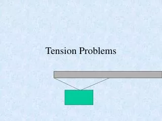
Tension hydropneumothorax
Tension hydropneumothorax. Air fluid level at right costophrenic angle Deeper right costophrenic angle as compared to the left Contralateral shift of mediastinum. ARDS. Bilateral diffuse fluffy infiltrates Normal cardiac size Tracheostomy tube


Tension hydropneumothorax
E N D
Presentation Transcript
Tension hydropneumothorax • Air fluid level at right costophrenic angle • Deeper right costophrenic angle as compared to the left • Contralateral shift of mediastinum
ARDS • Bilateral diffuse fluffy infiltrates • Normal cardiac size • Tracheostomy tube • Right subclavian central line going inside the right atrium • ECG wires
Right lung collapse - PA view • Ipsilateral shift of mediastinum and trachea • Bronchial cut-off sign suggestive of endobronchial obstruction • Rib crowding • Loss of volume • Obscured right mediastinal and cardiac outline • Obscured right hemidiaphragm (silhouette sign) • Compensatory hyperinflation of left lung • Prominent left pulmonary artery (cardiac output passing through single artery)
Right lung collapse – lateral view • Loss of gradually increasing transradiancy down the spine • Only one hemidiaphragm is visible (left) • Suspicion of mass in lower lobe with lymph node in mediastinum
Left lung collapse • Ipsilateral shift of trachea, carina and mediastinum • Bronchial cut-off sign (left mainstem bronchus) • Rib crowding • Loss of volume • Compensatory hyperinflation of right lung
Collapse with airbronchogram • Airbronchogram sign • Produced as a result of airspace opacification of the lung parenchyma • This results in visibility of the normally invisible black bronchi against a background of white opacification • Seen in consolidation and collapse with at least some patency of the bronchus
Left upper lobe collapse – PA view • Loss of volume on left side • Ipsilateral shift of trachea and mediastinum • Compensatory hyperinflation of left lung • Raised left hemidiaphragm (compare with right) with tenting • Haziness over the aortic knuckle (silhouette sign)
Left upper lobe collapse – Lateral view • Oblique fissure displaced anteriorly • Opacification anterior to the oblique fissure
Right upper lobe collapse - PA view • Loss of volume on right side • Opacification of right upper lobe • Transverse fissure raised • Right hilum is also raised
Right upper lobe collapse – lateral view • Oblique fissure displaced anteriorly • Transverse fissure pulled upwards • Opacification with loss of volume of right upper lobe
Left lower lobe collapse – PA view • Loss of volume on left side • Ipsilateral shift of the heart • Both hila are at the same level (left hilum has come down) • Double opacity behind the heart • Outline of left hemidiaphragm is obscured (silhouette sign) • Left hemidiaphragm is raised (watch the gastric bubble)
Left lower lobe collapse – Lateral view • Loss of gradually increasing transradiancy down the spine • One hemidiaphragm is clearly visible • Oblique fissure is displaced posteriorly
Miliary shadowing • Multiple small millet sized nodules throughout both lung fields
Mediastinal mass with left lower lobe collapse • Mediastinal widening in upper part mediastinum • Loss of volume on left side • Double opacity behind the heart • Left hemidiaphragm not visible • Heart shifted to the left side
Aspergilloma • Fungus ball with surrounding rim of air
Mesothelioma • Left sided pleural effusion • Associated lobulated pleural thickening • No shift of mediastinum due to encasement by mesothelioma
Pleural based mass • Cat under the rug appearance indicative of pleural based origin • Angle between chest wall and opacity is obtuse (>90o)
Sarcoidosis • Bilateral hilar lymphadenopathy • Right paratracheal strip enlargement • Bilateral infiltrates involving predominantly the mid zones
Pneumothorax • Left sided apical pneumothorax • Visceral pleural line is clearly visible • There should be no lung markings distal to the visceral pleural line
Effusion with collapse • Complete opacification of right hemithorax without significant contralateral shift of mediastinum • Absence of shift is indicative of concomitant collapse • Usually a sign of malignancy