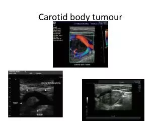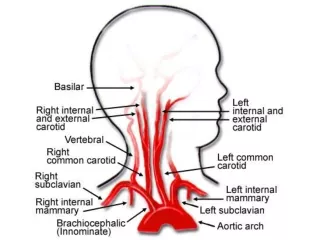
Carotid Dissection
Carotid Dissection . An Actual Case from : Detroit Medical Center, Harper University Hospital Vascular Lab Presented By : Angela Bowling Baker College Of Auburn Hills Vascular Ultrasound Winter, 2013. Overview. Carotid Dissection: Description Typical Symptoms Treatment Statistics

Carotid Dissection
E N D
Presentation Transcript
Carotid Dissection An Actual Case from: Detroit Medical Center, Harper University Hospital Vascular Lab Presented By : Angela Bowling Baker College Of Auburn Hills Vascular Ultrasound Winter, 2013
Overview • Carotid Dissection: • Description • Typical Symptoms • Treatment • Statistics • Patient Information: • Demographics • Symptoms • History • Test Ordered Images: • Waveform Interpretation • Hemodynamics • B-Mode • Preliminary Report • Conclusion
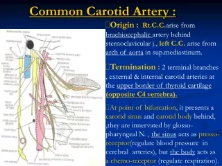
Carotid Dissection • “Hemorrhage within the carotid wall” • Trauma or Spontaneous • Intima tears • Dissecting the carotid artery into two parts • “True Lumen” • Usually crowded by false lumen • “False Lumen” • Can be patent or thrombosed • Waveforms typically to/fro flow in false lumen. Rumwell & McPharlin, (2009), Zwiebel & Pellerito, (2005). www.ispub.com, (2012
Carotid Dissection • Typical Symptoms: • Horner syndrome, (drooping eyelid, decreased pupil size) • “Neck pain, headache, tinnitus, transient monocular blindness” • Asymptomatic • Treatment: • Anticoagulation • Surgical Stent Zwiebel & Pellerito, (2005) Rumwell & McPharlin, (2009)
Carotid Dissection • RARE: In United States: only 2.6 per 100,000 patients. • How many in this room have scanned a Carotid Dissection? Detroit Medical Center, (2012), Zwiebel & Pellerito, (2005).
Patient Information • Initials: MJ • Sex: Female • Age: 80 years old • Symptoms: Syncope • History: • Prior Carotid Aneurysm with Corrective Surgery • Recent Fall down 2 steps due to syncope • Test Ordered: Bilateral Carotid Study Detroit Medical Center, (2012).
Rumwell & McPharlin, (2009), Detroit Medical Center, (2012), Zwiebel & Pellerito, (2005). Images: Waveforms .
Rumwell & McPharlin, (2009), Detroit Medical Center, (2012), Zwiebel & Pellerito, (2005). Images: Waveforms
Rumwell & McPharlin, (2009), Detroit Medical Center, (2012), Zwiebel & Pellerito, (2005). Images: Waveforms
Rumwell & McPharlin, (2009), Detroit Medical Center, (2012), Zwiebel & Pellerito, (2005). Images: Hemodynamics
Rumwell & McPharlin, (2009), Detroit Medical Center, (2012), Zwiebel & Pellerito, (2005). Images: Hemodynamics
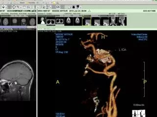
Images: B-Mode Rumwell & McPharlin, (2009), Detroit Medical Center, (2012), Zwiebel & Pellerito, (2005).
Preliminary Report Vascular Lab Report Indication: Syncope On the right: Smooth dense plaque visualized in ICA, PSV of ICA was 74 cm/sec ESV of 26cm/sec B-mode images and velocities are consistent with 1-39% diameter reduction of the proximal internal carotid artery. Right Brachial Systolic: 124 mmHg Left Brachial Systolic: 120 mmHg Vertebral arteries: appeared to have ante grade flow bilaterally On the left: Irregular dense plaque visualized in ICA, PSV of ICA was 107cm/sec ESV of 27cm/sec B-mode images and velocities are consistent with 1-39% diameter reduction of the proximal internal carotid artery. Detroit Medical Center, (2012)
Preliminary Report Continued On the left, there was a dissection in the mid common carotid artery visualized. The velocities pre dissection were 68 cm/sec, and the velocities post dissection were 228 cm/sec. There was a clip that was shadowing out a partial area of the common carotid artery. Doctor was notified of the results. Detroit Medical Center, (2012)
Detroit Medical Center, (2012) Conclusion • Patient: MJ, 80 year old female • Symptom: Syncope • History: Prior carotid aneurysm surgery Detroit Medical Center, (2012) Rumwell & McPharlin, (2009) Zwiebel and Pellerito, (2005)
Conclusion • Waveform Interpretation: Pre-dissection: • Laminar flow with spectral window intact • Moderate pulsatility • Normal PSV of 68 cm/sec False Lumen: • Reversal of flow : below baseline Post-dissection: • Turbulent flow with spectral window filled in • Moderate pulsatility • Suggested jet like PSV of 228 cm/sec • Hemodynamics: • Area of flow separation post dissection (Bernoulli Principle) • Turbulent flow post dissection with mosaic patterns • Retrograde flow in false lumen Detroit Medical Center, (2012) Zwiebel and Pellerito, (2005) Rumwell & McPharlin, (2009),
Detroit Medical Center, (2012) Conclusion • B-Mode Images: • Plaque visualized ICA Bilaterally • Left Mid CCA Dissection and clip with shadow • Suggested findings: • Left Side Common Carotid Artery Dissection, Clip shadowing area of dissection. • 1- 39% stenosis Bilaterally in ICA • Treatment: Anticoagulation
References References Internet Scientific Publications, ISPUB.com (2012) , The Internet Journal of Neurosurgery (Volume 7 No. 2), www.ISPUB.com Rumwell, C. & McPharlin, M. (2009). Vascular technology: An illustrated review (4thed.). Pasadena, CA: Davies Publishing Zwiebel, W. & Pellerito, J, (2005). Introduction to vascular ultrasonography (5thed.). Philadelphia, PA: Elsevier Saunders