Download
1 / 25
260 likes | 542 Vues
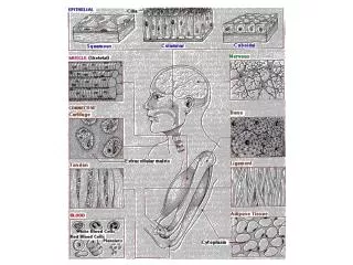
THE GROUND TISSUES EPITHELIAL TISSUES CONNECTIVE AND SUPPORTIVE TISSUES MUSCLE TISSUES NERVOUS TISSUE. HISTOLOGY 1.5.: CONNECTIVE AND SUPPORTIVE TISSUES. A diverse group of tissues that share a common origin from the mesenchyme of the embryo.

E N D
THE GROUND TISSUES EPITHELIAL TISSUES CONNECTIVE AND SUPPORTIVE TISSUES MUSCLE TISSUES NERVOUS TISSUE
HISTOLOGY 1.5.: CONNECTIVE AND SUPPORTIVE TISSUES • A diverse group of tissues that share a common origin from the • mesenchyme of the embryo. • They provide structural and metabolic support for other tissues and • organs throughout the body, bind the tissues to form organs, and the • organs to form organ systems and organism. • They contain blood vessels and mediate the exchange of nutrients, • metabolites and waste products between tissues and • the circulatory system. • THUS: • Origin: from the mesoderm • Blood supply: densely vascularized • Nerve supply: densely innervated
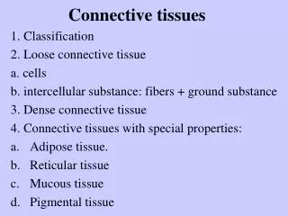
Tissue components of the connective and supportive tissues: • Cells of great variety (fixed and free cell types, see later) • Extracellular matrix (ECM) - ground substance • - fibers (collagen, elastic, reticular)
Extracellular matrix (ECM) I. THE GROUND SUBSTANCE • A highly hydrated gel in which the cells and fibers are embedded • Molecular components: • Proteoglycan consisting of dimericglycosaminoglycans (GAGs) • bound to a protein core: • chondroitin sulphate (cartilage, arteries, skin, cornea) • keratane sulphate (cartilage, bone, cornea) • heparane sulphate (arteries, lung) • dermatan sulphate (skin, tendon,ligaments, sclera, lung) • hyaluronic acid (vitreous humor of eye, synovial fluid, loose CT, • skin, cartilage, umbilical cord) • Hydrophilic side chains of GAGs bind a considerable amount • of extracellular fluid – water reservoir of the organism. • It is a part of the connective and supportive tissues not visible in • histological preparates, unless special staining methods are applied: • Toluidine-blue • PAS staining • Alcian-blue staining
Extracellular matrix (ECM) II. CONNECTIVE TISSUE FIBERS • COLLAGEN fibers • The most abundant protein in the body. • H.E-stained light micrograph Scanning electron micrograph Cross-striation observed in the transmission electron micro- scope Two isolated fibers artificially coloured in the computer (SEM)
Synthesis of collagen fibers: • Takes place in the rER of fibroblasts: • pro-a-chains form triple helices • These procollagen molecules are transferred to the Golgi complex • Tropocollagen molecules are released • via exocytosis from Golgi-vesicles • Tropocollagen molecules are assembled to form • collagen fibers in ECM Properties of collagen fibers • Non-branching bundles of changing thickness • High tensile strength • Poor sheer strength • They can be stretched to approximately 5 % of their initial length
Specific staining for the visualization of collagen fibers: Azan staining stains the collagen fibers selectively in blue colour other tissue componenst are stained red
TYPES OF COLLAGEN FIBERS Type I.: 2 a1 chains + 1 a2 chain the most common (bone, skin,tendon, ligaments) Type II.: 3 a1 chains predominates in cartilage Type III.: 3 a1 chains in embryonic CT and skin Type IV.: still unknown composition in the basal laminae of adults Type V.: still unknown composition mainly in the embryo
2. ELASTIC FIBERS They are present in organs whose normal function requires elasticity in addition to tensile strength Wall of the aorta stained with H.E. and resorcin fuchsin, specific staining for elastic fibres
SYNTHESIS OF ELASTIC FIBERS: • Takes place in the rER of fibroblasts (and smooth muscle cells): • tropoelastin molecules • Transformed to elastin, cross-linked and assembled in the • extracellular space • Microfibrils secreted prior to elastin provide scaffolding on which • elastin forms fibers and sheets • PROPERTIES OF ELASTIC FIBERS • Occur in the form of individual, branching and anastomosing fibers • They can be stretched 2.5 times of their original length • to which they return when released
3. RETICULAR FIBERS Individual collagen fibers coated by proteoglycans and glycoproteins. This coating causes their affinity for silver salts. They form delicate flexible networks around capillaries, muscle fibers, nerves, adipose cells and hepatocytes. Serve as scaffolding in endocrine, lymphatic and blood cell-forming organs. Reticular fibers around hepatocytes and around kidney tubules (as part of basement membrane)
CELL TYPES OF THE CONNECTIVE TISSUES • Fixed cells: fibrocytes • fibroblasts • mesenchymal cells • adipose cells • II. Free cells: macrophages • mast cells • plasma cells • melanocytes • blood-derived cells • (lymphocytes, monocytes, • eosinophils,neutrophils)
FIXED CELLS • I.1. Fibrocytes, fibroblasts: The most common fixed cell types. Fibroblast: metabolically more active (see before: synthesis of ECM components) (purple arrows). Fibrocyte: metabolically less active, more slender cell and nucleus, less cell organelles (green arrows)
FIXED CELLS • I.2. Mesenchymal cells • Pluripotent undifferentiated cells around blood vessels: • Serve as a reservoir of cells that can differentiate into other • connective tissue cell types.
I. FIXED CELLS: I.3. Adipose cells: LM SEM Adipose cells, single or in groups are normal components of the connective tissue (they may form a separate tissue: adipose tissue). Spherical or polyhedral cells with a large lipid droplet surrounded by a thin cytoplasmic rim. TEM
II. FREE CELLS: II.1. MACROPHAGES (histiocytes): in non-reactive CT are fixed, upon stimulation they become motile. Phagocytotic cell type, rich in hydrolytic enzymes. LM SEM TEM
THE MONONUCLEAR PHAGOCYTE SYSTEM Connective tissue: histiocytes Liver: Kupffer-cell Lung: alveolar macrophage Lymph nodes: free and fixed macrophages Spleen: free and fixed macrophages Bone marrow: fixed macrophages Serous cavities: pleural and peritoneal macrophages Bone: osteoclasts Central nervous system: microglial cells Skin: histiocytes Synovia: type A cells
II.FREE CELLS: II.2. Mast cells: around blood vessels in skin and intestine. TEM LM LM Large metachromatic granules: histamine, heparin, serotonin
II. FREE CELLS: II.3. Plasma cells: In the loose CT of gastrointestinal tract, respiratory system and female reproductive system, in the lymphatic tissue. They develop from B-lymphocytes, produce antibodies. LM TEM
II. FREE CELLS II.4. Melanocytes Large pigmented cells of neural crest-origin. They occur in the dermis of skin, meninges, choroid and iris.
II. FREE CELLS: II.5. Other free cells temporarily occurring in the CT Lymphocytes, monocytes, granulocytes (in details see the chapter „Blood” later)