Haemostasis
Haemostasis. Prof. K. Sivapalan. Thrombocytes. 2 – 4 μ m in diameter. Half life – 4 days. 300,000 / μ L. Break off from megakaryocytes. Colony stimulating factors and thrombopoietin- liver and kidney. 60 – 70 % in circulating blood - balance in spleen. Properties of Platelets.

Haemostasis
E N D
Presentation Transcript
Haemostasis Prof. K. Sivapalan
Thrombocytes • 2 – 4 μm in diameter. • Half life – 4 days. • 300,000 / μL. • Break off from megakaryocytes. • Colony stimulating factors and thrombopoietin- liver and kidney. • 60 – 70 % in circulating blood - balance in spleen. Haemostasis
Properties of Platelets • A ring of microtubules in periphery. • Extensively invaginated membrane. • Membrane contains receptors for: • Collagen, von Willebrand factor and fibrin. • Dense granules in cytoplasm: • Serotonin, ADP, other nuclear tides. • α – granules in cytoplasm : • Clotting factors and platelet-derived growth factor [PDGF – stimulates wound healing and mitogen for vascular smooth muscle.] Haemostasis
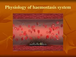
Platelet activation. • Binds to exposed collagen and von Willebrand factor (when damage to blood vessel). This is platelet adhesion. • This activates platelets [ADP] • Platelets change shape, put out psudopodia and release granules and causes platelet aggregation. Haemostasis
Changes in the platelet. • Platelet activation results in change in shape, putting out psseudopodia, release of granules and aehesion to other platelets. • Platelet Activating Factor secreted by neutrophils and monocytes stimulates G protein which activates phospholipase C to form diacylglycerol. This also causes release of granules. • Increased cytoplasmic calcium and diacylglycerol activate Phospholipase A2. This causes release of arachidonic acid from membrane phospholipids which is converted into Thromboxan A2 • Thromboxan and other substances released cause vasoconstriction, platelet aggregation, clot formation • Aspirin prevents the above reaction and alters the balance between thromboxan and prostacycline and prevents clotting in low doses. Haemostasis
Effects of platelet aggregation. • Repair of the blood vessels. • Block damaged capillaries. • Vasoconstriction. • Clotting. • [Test for platelet function: bleeding time.] Haemostasis
Thrombocytopenia. • Results in capillary bleeding [purpura]. • Caused by- • Marrow disorders. • Alcohol, cytotoxic drugs, viral infections. • Hereditary. • Immunologically mediated destruction. • Increased consumption of platelets. Haemostasis
Thrombocytosis. Causes: • Spleanectomy. • Postoperatively, delivery. • Haemorrhage or haemolysis. • Extreme exercise. Risk: • Thrombotic diseases- deep vein thrombosis. Haemostasis
Hemostasis. • Vascular spasm: local myogenic, serotonin – lasts for about 20 – 30 minutes. • Platelet plug. • Clotting of blood. • Organization by fibrous tissue. Haemostasis
Coagulation of blood – clotting. • Fibrinogen → Fibrin. • Polymerization of fibrin with branching. • Loose mesh of interlacing strands. • Formation of covalent bonds → dense, tight aggrigate. Haemostasis
Important reactions. Clot retraction. [platelets] 30 – 60 min. Stabilization. Fibrin. Fibrinogen Activated XIII Thrombin Platelet Factor, Ca++, Activated Factor V. Factor XIII Intrinsic system. 2 – 5 minutes. Factor x (activated) Extrinsic system. 15 – 30 Seconds. Prothrombin Haemostasis
Coagulation cascade. Tissue factor – Extrinsic system. [Tissue Thromboplastin- TPL+TFI] Contact with wettable, negatively charged surface – Intrinsic system. Activated VIII, Platelet factor (PL), Ca++ Ca++TPL+TFI Prekallikerin Kallikerin. HMW Kilinogen XII XIIa. XI XIIa IX IXa VIIa VII Ca++, PL, TPL X Xa Haemostasis
Role of liver in clotting. • Synthesizes: • Fibrinogen. • Prothrombin. • Other clotting factors. • Needs: • Vitamin K. • Removes activated clotting factors. Haemostasis
Propagation of clot formation. • Clot formation can be initiated at any vessel by damage to endothelium by platelet plug. • Activated clotting factors on the surface of the clot can cause further clotting. • Platelet plug can form on the surface of the clot which can initiate further clotting • Rapid flow wash off the factors which are diluted and removed in liver. Haemostasis
Clotting and Anti clotting mechanisms. • Clotting and anti clotting mechanisms are balanced under normal circumstances. • It is essential to maintain blood in liquid form but prevent loss if the vessels are damaged. Haemostasis
Anti clotting mechanisms. • Anti thrombin III [circulating protease] inactivates activated factors IX,X,XI and XII. Heparin facilitates it. • Prostacycline of Endothelium antagonizes thromboxane A2 of platelets. (aggregation) • Endothelium has thrombomodulin. It’s reaction with thrombin leads to fibrinolysis, inactivation of factor V and VIII. Haemostasis
Fibrinolytic system. Thrombomodulin in endothelium. Binds to Thrombin Prtotein C. Activated protein C.[APC] Inhibitor of tissue plasminogen activator inhibited. VIIIa Inactive VIIIa Va Inactive Va Plasmin. Plasminogen Lyses of fibrin. Tissue plasminogen activator Haemostasis
Abnormalities of clotting. Defective clotting: • Abnormalities of platelet function. • Congenital deficiency of clotting factors- • Hemophelia A – factor VIII [ X linked]. • Hemophelia B – factor IX. • Von Willebrand factor deficiency. • Vitamin K deficiency and Liver diseases. Enhanced clotting: • Increased platelets. • Absent Protein C. Intravascular Clotting is thrombosis and if carried in blood it is embolism. Both can obstruct blood vessels and cause ischemia to organs. Haemostasis
Anticoagulants. • Heparin promotes antithrombin III which inactivates factors IX, X, XI and XII. • Warfarin inhibits Vitamin K. • Streptokinase activates plasminogen and disolves fibrin. [snake, bacteria] • Aspirin reduces thromboxan A2 formation. Haemostasis
Investigations. • Clotting time. • Allow blood to clot and record time. • Prothrombin time. • Anticoagulated blood and prothrombin. Add calcium chloride and record time. • Bleeding time. • Time taken to stop bleeding. Haemostasis