Download
1 / 34
350 likes | 420 Vues
Explore the structure and function of the human body, from cells to organ systems. Learn about the hierarchy of organizational levels and the major functions of different body systems. Discover anatomical terminology for accurate understanding.

E N D
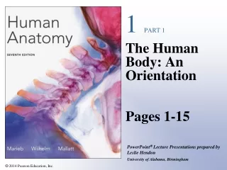
1 PART 1 The Human Body: An Orientation Pages 1-15
An Overview of Anatomy Anatomy The study of the structure of the human body Physiology The study of body function
The Hierarchy of Structural Organization Chemical level—atoms form molecules Cellular level—cells and their functional subunits Tissue level—a group of cells performing a common function
The Hierarchy of Structural Organization Organ level—a discrete structure made up of more than one tissue Organ system—organs working together for a common purpose Organism level—the result of all simpler levels working in unison
Figure 1.1 Recognizing connections between structural levels leads to better understanding of organismal function.
Figure 1.2a-f The body’s organ systems and their major functions. Skeletalmuscles Hair Nails Skin Bones Joint Muscular System Integumentary System Skeletal System Allows manipulation of the environment,locomotion, and facial expression.Maintains posture and produces heat. Forms the external body covering and protectsdeeper tissues from injury. Synthesizes vitaminD and houses cutaneous receptors (pain,pressure, etc.) and sweat and oil glands. Protects and supports body organs andprovides a framework the muscles useto cause movement. Blood cells areformed within bones. Bones store minerals. Pineal gland Brain Pituitarygland Thyroidgland Heart Thymus Adrenalgland Pancreas Testis Nerves Ovary Bloodvessels Spinalcord Cardiovascular System Nervous System Endocrine System Blood vessels transport blood, which carriesoxygen, carbon dioxide, nutrients, wastes, etc.The heart pumps blood. Glands secrete hormones that regulate processessuch as growth, reproduction, and nutrient use(metabolism) by body cells. As the fast-acting control system of the body, itresponds to internal and external changes byactivating appropriate muscles and glands.
Figure 1.2g-l The body’s organ systems and their major functions. Nasalcavity Oral cavity Red bone marrow Esophagus Pharynx Thymus Bronchus Larynx Lymphaticvessels Trachea Thoracicduct Liver Lung Stomach Smallintestine Spleen Largeintestine Lymph nodes Rectum Anus Lymphatic System/Immunity Respiratory System Digestive System Picks up fluid leaked from blood vesselsand returns it to blood. Disposes of debrisin the lymphatic stream. Houses white bloodcells (lymphocytes) involved in immunity.The immune response mounts the attackagainst foreign substances within the body. Keeps blood constantly supplied withoxygen and removes carbon dioxide.The gaseous exchanges occur throughthe walls of the air sacs of the lungs. Breaks down food into absorbable unitsthat enter the blood for distribution tobody cells. Indigestible foodstuffs areeliminated as feces. Mammaryglands (inbreasts) Kidney Prostategland Ureter Ovary Penis Ductusdeferens Urinarybladder Testis Uterinetube Uterus Scrotum Urethra Vagina Male Reproductive System Female Reproductive System Urinary System Overall function is production of offspring. Testes produce sperm and male sex hormone, and maleducts and glands aid in delivery of sperm to the female reproductive tract. Ovaries produce eggsand female sex hormones. The remaining female structures serve as sites for fertilization anddevelopment of the fetus. Mammary glands of female breasts produce milk to nourish the newborn. Eliminates nitrogenous wastes from thebody. Regulates water, electrolyte, andacid-base balance of the blood.
Systemic v. Regional Anatomy Systemic—study of anatomy by system Regional—study of anatomy by region Most students use a combination of regional and systemic study
Scale: Length, Volume, and Weight Metric system provides a precise system of measurement
Anatomical Terminology Anatomical position—a common visual reference point Person stands erect with feet together and eyes forward Palms face anteriorly with the thumbs pointed away from the body Directional terminology—refers to the body in anatomical position Standardized terms of directions are paired terms
Figure 1.3a Anatomical position and regional terms. Axial region Cephalic (head) Frontal Orbital Nasal Upper limb Oral Acromial Mental Brachial (arm) Cervical (neck) Antecubital Thoracic Antebrachial (forearm) Sternal Axillary Carpal (wrist) Mammary Abdominal Umbilical Manus (hand) Pelvic Pollex Palmar Inguinal (groin) Digital Lower limb Coxal (hip) Pubic (genital) Femoral (thigh) Patellar Crural (leg) Fibular or peroneal Pedal (foot) Thorax Tarsal (ankle) Abdomen Metatarsal Digital Back (Dorsum) Hallux Anterior/Ventral
Figure 1.3b Anatomical position and regional terms. Appendicularregion Cephalic Otic Occipital (back of head) Upper limb Acromial Cervical Brachial (arm) Olecranal Antebrachial (forearm) Back (dorsal) Scapular Vertebral Lumbar Manus (hand) Sacral Metacarpal Digital Gluteal Perineal(between anusand externalgenitalia) Lower limb Femoral (thigh) Popliteal Sural (calf) Fibular or peroneal Thorax Abdomen Pedal (foot) Back (Dorsum) Calcaneal Plantar Posterior/Dorsal
Anatomical Terminology Directional terms Regional terms—names of specific body areas Axial region—the main axis of the body Appendicular region—the limbs
Body Planes and Sections Coronal (frontal) plane Lies vertically and divides body into anterior and posterior parts Median (midsagittal) plane Specific sagittal plane that lies vertically in the midline
Body Planes and Sections Transverse plane Runs horizontally and divides body into superior and inferior parts
Figure 1.4 Planes of the body with corresponding magnetic resonance imaging (MRI) scans. Median section(midsagittal) Frontal plane Median(midsagittal)plane Transverseplane Rectum Intestines Vertebralcolumn Frontal section(through torso) Transverse section(through torso, inferior view) Spleen Liver Subcutaneousfat layer Liver Stomach Pancreas Spinal cord Aorta Left andright lungs Heart Arm Body wall
Body Cavities and Membranes Dorsal body cavity Cranial cavity Vertebral cavity
Body Cavities and Membranes Ventral body cavity Thoracic cavity—divided into three parts Two lateral parts each containing a lung surrounded by a pleural cavity Mediastinum—contains the heart surrounded by the pericardial sac
Body Cavities and Membranes Ventral body cavity (continued) Abdominopelvic cavity—divided into two parts Abdominal cavity—contains the liver, stomach, kidneys, and other organs Pelvic cavity—contains the bladder, some reproductive organs, and rectum
Body Cavities and Membranes Cranial cavity (contains brain Dorsal body cavity Thoracic cavity (contains heart and lungs) Vertebral cavity (contains spinal cord) Diaphragm Abdominal cavity (contains digestive viscera) Pelvic cavity (contains urinary bladder, reproductive organs, and rectum) Dorsal body cavity Ventral body cavity (a) Lateral view Figure 1.6a
Body Cavities and Membranes Cranial cavity Dorsal body cavity Ventral body cavity Vertebral cavity Superior mediastinum Thoracic cavity (contains heart and lungs) Pleural cavity Pericardial cavity within the mediastinum Ventral body cavity (thoracic and abdominopelvic cavities) Diaphragm Abdominal cavity (contains digestive viscera) Abdomino- pelvic cavity Pelvic cavity (contains urinary bladder, reproductive organs, and rectum) (b) Anterior view Figure 1.6b
Body Cavities and Membranes Serous cavities—a slit-like space lined by a serous membrane Pleura, pericardium, and peritoneum Parietal serosa—outer wall of the cavity Visceral serosa covers the visceral organs
Body Cavities and Membranes Lung Ribs Parietal pleura Pleural cavity with serous fluid Visceral pleura Diaphragm (a) Serosae associated with the lungs: pleura Figure 1.7a
Body Cavities and Membranes Heart Parietal pericardium Pericardial cavity with serous fluid Visceral pericardium (b) Serosae associated with the heart: pericardium Figure 1.7b
Body Cavities and Membranes Visceral peritoneum Anterior Peritoneal cavity (with serous fluid) Liver Stomach Parietal peritoneum Kidney (retroperitoneal) Wall of body trunk Posterior (c) Serosae associated with the abdominal viscera: peritoneum Figure 1.7c
Body Cavities and Membranes Outer balloon wall (comparable to parietal serosa) Air (comparable to serous cavity) Inner balloon wall (comparable to visceral serosa) (d) Model of the serous membranes and serous cavity Figure 1.7d
Abdominal Quadrants Abdominal quadrants divide the abdomen into four quadrants Right upper and left upper quadrants Right lower and left lower quadrants
Figure 1.8 Abdominal quadrants. Diaphragm Liver Spleen Stomach Gallbladder Left upperquadrant(LUQ) Right upperquadrant(RUQ) Transverse colonof large intestine Ascending colonof large intestine Descending colonof large intestine Small intestine Cecum Right lowerquadrant(RLQ) Left lowerquadrant(LLQ) Initial part ofsigmoid colon Appendix Urinary bladder Anterior view of the four quadrants showing thesuperficial organs The four abdominopelvic quadrants
Microscopic Anatomy Microscopy—examining small structures through a microscope Light microscopy illuminates tissue with a beam of light (lower magnification) Electron microscopy uses beams of electrons (higher magnification)
Microscopic Anatomy Cytoplasm Cell nuclei Extracellular material (a) Light micrograph (330) (c) Scanning electron micrograph, artificially colored (2900) (b) Transmission electron micrograph, artificially colored (870) Figure 1.9a–c
Microscopic Anatomy Preparing human tissue for microscopy Specimen is fixed (preserved) and sectioned Specimen is stained to distinguish anatomical structures Acidic stain—negatively charged dye molecules Basic stain—positively charged dye molecules
Microscopic Anatomy Scanning electron microscopy Heavy metal salt stain—deflects electrons in the beam to different extents Artifacts Minor distortions of preserved tissues Not exactly like living tissues and organs