The Sensory Organs
670 likes | 727 Vues
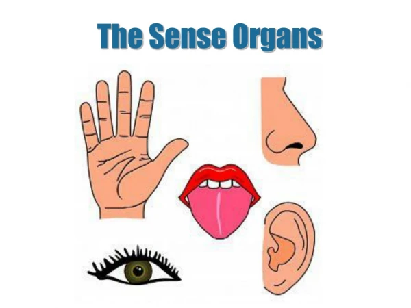
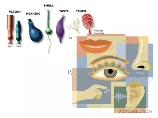
The Sensory Organs. Pg. 213-. The Eye. The eye is the sensory organ related to vision It picks up light rays given off by light sources or reflected by objects A normal eye can differentiate among 2000 or so colours, and can adapt to light intensity

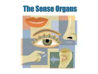
The Sensory Organs
E N D
Presentation Transcript
The Sensory Organs Pg. 213-
The Eye • The eye is the sensory organ related to vision • It picks up light rays given off by light sources or reflected by objects • A normal eye can differentiate among 2000 or so colours, and can adapt to light intensity • Take a look at Fig 7.19 and look at all of the parts of the eye
Sclera • Rigid, opaque membrane that protects the eye from shock and gives it shape • It is also called the White of the eye
Choroid • Middle layer of the eye, with blood vessels that nourish the eye • Think like you are peeling an orange (all the layers you would cross are in the eye)
Retina • Innermost layer at the back of the eye, thin and beige • Covered with millions of light-sensitive nerve cells that transform incoming data into nerve impulses • The junction point between the optic nerve and retina, the BLIND SPOT, has NO sensitivity to light
Cornea • Clear and rigid membrane that is an extension of the sclera in front • It is slightly more dome-shaped than the rest of the eye
Iris • An extension of the choroid, this pigmented (coloured) membrane has an opening, the PUPIL, in the centre, which serves to regulate the amount of light coming into the eye • The more light, the smaller (contracted) the pupil becomes • The less light, the larger (dilated) the pupil becomes • Can you see better when you squint? Why?
Lens • Flattened sphere that focuses light rays on the retina • It is held in place by muscles, which flatten it or make it more spherical, thus changing the focus
Aqueous humour • Transparent liquid that fills the space between the cornea and the lens • Aqueous is in the front of the eye (A is first in the alphabet) • Between the cornea and the lens
Vitreous humour • Transparent jelly-like substance that fills the space between the lens and the retina
Nerve cells in the Retina • The nerve cells in the retina are photo-receptors • Meaning they convert light into nerve impulses • Some of the nerve cells are called Cones • Only able to distinguish colours • While other are called Rods • Only able to distinguish variations in light intensity
Messages sent to brain • Nerve impulses from retina cells are sent to the brain through the optic nerve • Info is then processed and analyzed by the brain • The brain superimposes (copies) the image it received from each eye • This is how we are able to estimate distances and objects contours (shapes)
EYE didn’t know that! Answers to your questions • Iris color is a highly complex phenomenon consisting of the combined effects of texture, pigmentation, fibrous tissue and blood vessels within the iris • The color of your eyes depends on your genes, which you inherit from your parents. Eye color comes from a combination of a black and a yellow pigment called melanin in the iris of your eye. An increasing proportion of the yellow melanin, in combination with the black melanin, results in shades of colors between brown and blue, including green and hazel.
EYE didn’t know that! Answers to your questions • Eyes can change color due to age or disease – most usually fading but sometimes darkening. If you notice your eyes losing color its time to visit your doctor. • Babies eyes change from blue to their natural color by the age of three. This is due to exposure to light which triggers the production of melanin in the iris.
EYE didn’t know that! Answers to your questions • Some people have one brown eye and one green eye. This abnormality can be caused by either a trauma in the womb, faulty developmental pigment transport or a benign genetic disorder. • Human albinos generally have very light blue eyes, as the un-pigmented color of the human iris is a pale blue
EYE didn’t know that! • The pupil appears black because of the layer of black pigmented cells that line the back of the retina and absorb the light. • Cat pupils:
In low light levels the cat’s pupil must be able to open as wide as possible, but also be able to contract to a very small size to protect the sensitive retina in bright sunlight. In human eyes, this size variation of the pupil is controlled by a circular ciliary muscle, but this limits the amount of size variation. In cats however, the same process is controlled by two, shutter-like ciliary muscles, which gives the cat it’s characteristic slit-like pupil in bright light conditions.
Lens accommodation cont’ • Light refracts when it hits the lens. • We have a biconvex lens that allows light to converge to one point.
Lens accom. • When you look at a distant object: • Light rays are parallel • Lens does not have to change shape • Rays converge and focus on retina • When you look at something close up: • Light rays diverge (move away from one another) • Lens has to become more curved to make the light rays converge and focus on the retina. • Ciliary muscles contract and pull the zonular ligaments attached to the lens.
Eye problems • Emmetropia = normal eyes, proper functioning. • Light rays focus on the retina and the brain has a clear image to analyze.
Eye probs cont. • Myopia = near-sightedness • You can see close up but not far away • Can happen because of an elongated eyeball or an overly curved lens. • Light rays converge too soon and the image ends up in front of the retina. • Fix it with a concave (diverging) lens
Eye probs cont… • Hypermetropia = far-sightedness • Sees distant objects very well but not objects close up • Because of a shortened eyeball or a insufficiently curved lens • The image is focused behind the retina. • Corrected with a biconvex lens.
Eye probs cont. • Presbyopia: like hypermetropia • Makes nearby objects hard to see • Because of a loss of lens flexibility as you get older • Fix using biconvex lens
The ear is the receptor organ of sound3 parts: • Outer ear • Middle ear • Inner ear
The Ear pg 215 • The ear is the sensory organ associated with hearing • It picks up sound and converts them into nerve impulses • The ear is divided into 3 sections • The outer ear • The middle ear • The inner ear • Look at Fig 7.24
Outer Ear • Pinna (auricle): only visible part of the ear. It is shaped like a funnel to pick up sound vibrations easily from air • Auditory canal: slightly curved, about 2.5 cm long canal that carries sound vibrations to the eardrum. It is lined with fine HAIR and sebaceous glands (produce wax)= prevent foreign bodies from entering the ear
Middle Ear Fig 7.25 • Tympanic membrane (eardrum): thin, flexible and fibrous membrane about 1 cm in diameter, which moves to the rhythm of sound wave vibrations • Ossicles (bones): miniature bones located in the temporal bone (think Temple). 3 bones= hammer, anvil, stirrup – can move in relation to one another • Eustachian tube: canal that links the middle ear to the pharynx. It equalizes the pressure on either side of the eardrum during swallowing (like on an airplane)
Inner Ear • Semi-circular canals: canals that form a liquid-filled maze in temporal bone. They regulate balance when the body is in motion • Vestibule: liquid-filled structure that links the semi-circular canals to the cochlea. The vestibule plays a role in balancing the body in a static position • Cochlea: liquid-filled structure, whose walls are covered with auditory nerve cells linked to the auditory nerve • See a pattern???
Stimulus -> Pinna -> Auditory canal -> Eardrum -> Hammer -> Anvil -> Stirrup -> Vestibule ->Cochlea -> Cilia -> Nerve cells -> Auditory nerve -> Temporal lobe • See p. 213-214 in textbook for picture and explanation
Sound • Sound is created by vibrations, usually in the air • Sources of sound (voices, music, motors, etc) cause changes in air pressure, which create sound waves • Sound waves are channeled by the Pinna into the ear • Through its various structures • Until they reach the fluid-filled cochlea • See Fig 7.26 show the path perfectly… LOOK!
Creating sound…. • The walls of the cochlea are lined with nerve cells • Their endings are sensitive to the vibrations in the liquid cochlea • These cells transform the info they receive into nerve impulses • This then travel to the cerebrum thru auditory nerves • The brain analyzes this and we ‘hear’ sound
Balance • The ears also play an important role in balance • Inner ear continuously monitors the position of the head • The constant monitoring can, for example, help a diver find the water’s surface • The cells also help keep your balance when you are not moving (stabilize our posture) • (the ears and eyes work together in this…stand up and close your eyes and see what happens)
Balance in motion • Other cells in the inner ear maintain balance when our bodies are moving • They allow us to detect changes in speed and direction • Helping us walk and dance without falling over
The Skin • The skin is the sensory associated with touch • It is a very large organ as it covers the entire surface of the body
Epidermis • Your epidermis is showing!!! • Dead layer: outer skin layer. Atmospheric pressure causes the cells to burst and die • Living layer: Layer of constantly dividing cells. New cells push old cells to the surface. They help in the healing process
Dermis • Sensory receptors: structures that pick up stimuli • Blood vessels: vessels that nourish the skin cells • Sebaceous glands: glands that secrete sebum, an oily substance that waterproof the skin • Sweat glands: glands that produce sweat, which is carried to the skin surface thru pores • Hair: structures arising from the dermis and partially covering the epidermis. An adjacent muscle can contract, making hair stand erect producing goose bumps!
Sensory receptors in skin… • Allow us to experience the sensation of: • Tactile sensations (touch, pressure) • Thermal sensation (heat, cold) • Painful sensation (pain) • Each sensation is detected by receptors whose nerve endings are free or contained in a protective capsule • See Fig 7.31
Sensory receptors • Sensory receptors are not spread out equally over the body • Some surface areas, like the underside of your wrist or your cheeks, are more sensitive to heat • But places like soles of our feet have more receptors related to touch or pressure • Sensory receptors combine to form sensory nerves that transport info to the cerebrum
Skin … • The skin is not only associated with touch, it also protects the body’s internal organs and block foreign bodies from entering our body • Also helps eliminate waste by sweating • And helps produce Vit D, which is needed by the body to absorb calcium • See Fig 7.32 to see where in the brain is related to skin receptors