Download
1 / 50
540 likes | 859 Vues
Idiopathic Scoliosis . dr n. med. Dariusz Mątewski. Lateral deviation of spine – postural deformity or scoliosis?.

E N D
Idiopathic Scoliosis dr n. med. Dariusz Mątewski
Lateral deviation of spine – postural deformity or scoliosis? • Postural deformityof body (sensu stricte) is the clear deviation from the normal posture, which is possible actively and passively corrected orcan disappearduring normal growth of body
Lateral deviation of spine – postural deformity or scoliosis? • The most frequent deformity of posture is a lateral curvature of the spine in the thoracolumbar section, mostly in left direction.
Lateral deviation of spine – postural deformity or scoliosis? • Idiopathic scoliosis – developmental, three-plane deformity of spine and trunk of unknown etiology • Frontal plane – lateral deviation • Sagittal plane – disturbance of physiological lordosis and kyphosis • Horizontal plane – axial rotation of vertebras
Scoliosis - definition • Scoliosis – spine curvature with Cobb angle > 10°
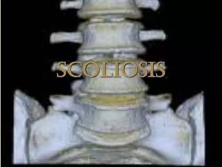
Anatomy • All bony elements are altered • Vertebra are wedge shaped • Rib vertebral angle altered • Pedicles rotated • Discs are wedged as well
Terminology • Named by apex • Cervical if between C2-C6 • Cervicothoracic if between C7-T1 • Thoracic if between T2-T11 • Thoracolumbar if between T12-L1 • Lumbar if between L2 and below • Primary vs secondary • Structural vs non-structural
Types of Scoliosis • Congenital • Muscular • Duchenne atrophy • Neuromuscular • Cerebral palsy • neurofibromatosis • Syndrome related • Marfan’s syndrome • Idiopathic • 80% are this
Etiological Theories • Genetic • Tissue deficiencies • Growth abnormalities • Central nervous system alteration
Genetic • 11% incidence in first relatives of patients • Normal incidence < 3% • Monozygote twins more common • No gene identified to date
Tissue Deficiencies • Marfan’s syndrome deficient fibrillin • Osteopenia noted in girls • Elevated calmodulin • Involved in contractile properties thru actin & myosin • Elevated in platelets • No consistent findings to date
Growth Abnormality • Asymmetrical vertebral growth • Hueter-Volkman effect is suppression of growth on concave side • Hypokyphosis during growth spurt • No increased incidence with growth hormone • No initiating factor identified
Central Nervous System • Different size cerebral cortices • Altered equilibrium • Primary or secondary • Deficient melatonin • Chicken model • Inconclusive in humans
Classification • Infantile: 0-3 years old (.5%) • Juvenile: 4-11 years old (10.5%) • Adolescent: 10-17 years old (89%) • Adult: >18 years old
Lenke classification • Typ 1- skolioza jednołukowa piersiowa (MT) • Typ 2- skolioza podwójna piersiowa (DT) • Typ 3- skolioza podwójna piersiowa i lędźwiowa z przewagą piersiowej (DM) • Typ 4- skolioza trójłukowa (TM) • Typ5- skolioza piersiowo-lędźwiowa jednołukowa albo lędźwiowa jednołukowa (TL/L) • Typ 6- skolioza podwójna z przewagą lędźwiowej lub piersiowo- lędźwiowej (TL/L structural MT)
Gruca/Wiesflog classification • I grade scoliosis - <20° according to Cobb angle • I grade scoliosis - <20-40° according to Cobb angle • III grade skoliosis - >40° wg according to Cobb angle
History • Family history • Affected sibling 7 times more frequent • Affected parent 3 times more frequent • Recent growth history • Sexual maturity • Pain • ‘Fatigue pain’ • Post diagnostic pain • ‘Severe pain’
Physical Exam • Iliac crest height • Leg length discrepancy • Shoulder height • Arm trunk space • Scapular position • Trunk shift • Inspection of skin • Café au lait spots
Neurologic Exam • Observe gait • Hop test • Heel and toe walk • Reflexes
Imaging • Plain x-rays • Need standing 36 inch cassette • Posterior to anterior • Decrease thyroid and breast exposure 3-7 fold • Note rotation • Measure deformity by Cobb method • Skeletal maturity
Rotation • Spinous process rotates into concavity • Pedicle position
Skeletal Maturity • Gruelich & Pyle atlas • Triradiate cartilage fusion • Risser sign
MRI • Neurologic deficit • Infantile and juvenile curves • Spinal cord abnormality in younger children • Infantile idiopathic scoliosis 50% • Juvenile 20%
Infantile Treatment • Must prove idiopathic • 90% are left thoracic • 3 female : 2 male • 90% resolve spontaneously • Predict progression by RVAD • < 20 degrees 83% resolve • >20 degrees 84% progress
Juvenile Treatment • Younger onset likely to progress • >30 degree curve almost always progress • Some adolescent curves are missed juvenile
Adolescent Treatment • Most curves <10 degrees • Boys = girls for these curves • Usually don’t progress • More sever curves (>30 degrees) • 8 girls : 1 boy • Predicting who will progress
Risk for Progression • Younger onset • Skeletal age • Risser 0-1 at presentation 60-70% progress • Risser 3 only 10% risk • Menses starts after growth spurt • Female more likely than male • Curve pattern • Apex above T12 • Degree at presentation • 20-29 degrees 68% risk for progression • 30-59 degrees 90% risk for progression
Natural History • If curve <30 degrees at maturity • No adult consequences • Unlikely to ever progress • Curves >45 degrees may progress a degree/year • Mortality not increased unless curve >90 degree • Right heart failure • Decreased pulmonary function
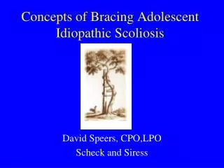
Non-Operative Treatment • <25 degrees monitor every 4-12 months • Depends on skeletal maturity • >25 degrees monitor every 3-6 months • >30 degrees in skeletally immature brace • Curve change by 10 degrees brace • Curve >40-45 degrees surgery
Bracing • Duration and time in brace • 23 hours per day • Wear until skeletally mature • Types • Milwaukee • Underarm orthosis • Charleston night time bending brace • Electrical stimulation
Successful Bracing • Prevent curve progression • Randomized study • Braced 74% did not progress • Not braced 34% did not progress • Electrical stimulation • 33% did not progress • Charleston brace still controversial
Problems with Braces • Argued efficacy • Narrow treatment window to initiate • Poor compliance • Must have good orthotist • Curves corrected by 20 degrees in brace do better
Surgery • Failed bracing • Curves >45 degrees • Unbalanced curves >40 degrees • Surgery is fusion with instrumentation
Surgical treatment • Technique of choice is transpedicular instrumentation with hooks and rods combine with posterior fusion according to CD rules
Preoperative planning • Identification of stable vertebras
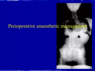
Surgical technique – patient positioning • Pronate position • General anesthesia • Antybiotic prophylaxis • Blood supply
Surgical technique – approach • Incision in posterior midline
Surgical technique – approach • Periostal incision,
Surgical technique • Transpedicular screw implantation