Stroke: Understanding Management for EMS
Stroke: Understanding Management for EMS. Silver Cross EMS System Adapted from and thanks to Alameda EMS System Additional material from Erika Ball, RN, BSN. Stroke Management for the EMS Provider. At the completion of this module, the EMS Provider will be able to:

Stroke: Understanding Management for EMS
E N D
Presentation Transcript
Stroke: Understanding Management for EMS Silver Cross EMS System Adapted from and thanks to Alameda EMS System Additional material from Erika Ball, RN, BSN
Stroke Management for the EMS Provider At the completion of this module, the EMS Provider will be able to: Describe the various types of stroke and their etiology. Discuss the imperatives for best practice in regard to EMS stroke management. List 5 or more risk factors for acute stroke. Define “penumbra” and how this concept is important in stroke. Generally describe the major vessels involved in acute ischemic stroke. Discuss the “therapeutic window” for thrombolytic therapy in stroke. Identify interventions that individual EMS providers can make to improve outcomes in stroke.
Is STROKE a health problem in the US today? • 700,000 strokes every year • Stroke is the 3rd leading cause of death • One person dies of stroke every 3 minutes • Stroke is the leading cause of serious, long term disability • 5 million stroke survivors, but with substantial morbidity: • 18% unable to return to work • 4% require total custodial care
Is STROKE a health problem in the US today? • Only 50-70% of stroke survivors regain functional independence • 20% are institutionalized within 3 months • 22% of men & 25% of women die within 1 year of their first stroke • Locally, African-Americans have 50% more strokes than Caucasians, and twice as many as Asians and Hispanics (Statistics from the American Stroke Association)
Women & Stroke • Stroke kills more than twice as many American women every year as breast cancer • More women than men die from stroke • Women over age 30 who smoke and take high-estrogen oral contraceptives have a stroke risk 22 times higher than average (National Stroke Association)
Is STROKE a health problem in the US today? • YES, stroke is a major health problem in the US today. • EMS Providers are closely involved with this patient population and are a vital component of the “Stroke Chain of Survival”. • Increased knowledge and personal motivation on the part of EMS providers can: • Greatly reduce death and disability due to stroke. • Improve stroke centers’ ability to provide thrombolytic therapy. • Make a positive impact on communities’ strides to reduce costs for healthcare and improve outcomes.
Goals for EMS Provider Care of Stroke Patients • Improve knowledge of identification of stroke signs and symptoms. • Develop a rapid assessment process. • Facilitate transfer of stroke victims to Primary Stroke Centers in the quickest and safest manner. • Pre-notify the Stroke Center, “Possible acute stroke in route.” • Encourage family members familiar with the patient care to either ride with the transfer vehicle or drive to the stroke center ASAP to provide more patient information.
Goals for EMS Provider Care of Stroke Patients • Obtain reliable list of meds taken or bring bag of all medications taken. • Obtain a set of vital signs and finger stick blood sugar at the site. • Reliably identify family’s best estimation of when the patient was “last seen normal”. • Administer the Cincinnati Pre-hospital Stroke Scale. • Provide the receiving facility with a quick, complete verbal report that incorporates the information obtained since arrival on scene.
Review: Anatomy & Physiology of Acute Ischemic Stroke • What is acute ischemic stroke? • What is the major vasculature involved? • When circulation is suddenly reduced, how quickly is brain tissue affected? • What is “penumbra”? • What are the types and etiologies of stroke? • What about different stroke symptoms?
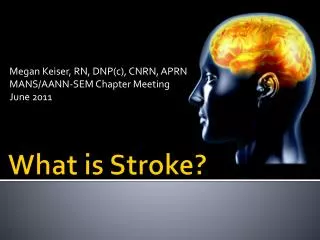
What Is Stroke ? A stroke occurs when blood flow to the brain is interrupted by a blocked or burst blood vessel.
One quarter of cardiac • output goes to the 5-6 • pound organ—the brain. • The brain needs a • constant supply of: • Oxygen • Glucose • Other nutrients • Circulation is supplied • via 2 pairs of arteries: • Internal carotids • Vertebrals
PENUMBRA (That tissue surrounding the infarct that is salvageable, but at risk.) Rapid transfer to the stroke center will allow for protection of penumbra through emergency interventions and medical management.
Cerebrovascular Disease: Pathogenesis Ischemic Stroke (83%) Hemorrhagic Stroke (17%) Atherothrombotic Cerebrovascular Disease (20%) Intracerebral Hemorrhage (59%) Cryptogenic (30%) Subarachnoid Hemorrhage (41%) Lacunar (25%) Small vessel disease Embolism (20%) Albers GW, et al. Chest. 1998;114:683S-698S. Rosamond WD, et al. Stroke. 1999;30:736-743.
Acute Ischemic Stroke(What do you see?) • Deficits: • Unilateral (though not always) weakness • Unilateral sensory deficit • Visual deficits (blindness, gaze palsy, double) • Speech (slurred – a motor dysfunction) • Language (aphasia – damage to the brain’s speech center) • Ataxia (lack of coordinated movement) • Cognitive impairment • Like real estate—Location, Location, Location
What Are the Effects of Stroke? • Left Brain
What Are the Effects of Stroke? • Right Brain
Stroke Assessment Scale(Cincinnati Pre-hospital Stroke Scale) “The sky is blue in Cincinnati.” Any abnormality means an abnormal Cincinnati scale for stroke. Probably accurately detects stroke 80% of the time.
Stroke Assessment in the Field • Administer Cincinnati Scale. • Code 38 of the SMO’s: Suspected Stroke • If abnormal, facilitate a rapid transfer to the primary stroke center. • Pre-notify the receiving stroke center—”possible acute stroke in route”.
Identify Time “Last Seen Normal” • A 75 year old man with HTN and diabetes finishes dinner with a friend at 8pm. He drives himself the short distance home that night, and a daughter stops by the next morning to find him still in bed and with right side weakness and severe aphasia. When do we assume the stoke occurred? (Answer: “last known normal at 8pm) • A 35 year old hypertensive man who is known to be non-compliant with meds is found slumped over in his car in a job site parking area at 3pm. In the ED he was found to have a massive left hemispheric ischemic stroke. His wife said he left for work at 7am that morning as normal, and she had a clear and normal cell phone conversation with him at 12:30pm. At 1pm a co-worker stated the man said he wasn’t feeling well and was going to his car to rest. At the time the co-worker noticed his speech was slurred. What time can we use as the time “last known normal”? (Answer: 12:30pm)
Types of Acute Ischemic Strokes • Middle Cerebral Artery Stroke • Vertebral—Basilar Artery Strokes • Lacunar Strokes
CT Scan of Acute Ischemic Stroke (Left MCA territory stroke)
Types of Strokes(Middle Cerebral Artery – MCA) • The most common artery occluded in AIS—can be proximal or from carotid circulation. • Features: • Motor/Sensory Deficit: face, arm, leg • Speech deficit – dysarthria (slurred speech) • Language deficit – if in dominant hemisphere • Gaze palsy – eyes directed towards side of AIS • Blindness – visual field cut (homonymous hemianopsia)
Types of Strokes(Vertebral—Basilar Artery) • Features: • Cranial nerve involvement – hearing, visual, facial, swallowing • Can have bilateral weakness • Cerebellar signs – ataxia • Sensory deficits • Vertigo – often nystagmus • Nausea and vomiting • Common to have waxing and waning symptoms
Lacunar Strokes These strokes are ischemic in nature. Mainly caused by HTN. Occurs in the small penetrating arteries of the brain. Presentation – affects the arm, leg, and face, sometimes silent. Deficits are equal to all areas.
Conditions That Mimic AIS • Bell’s Palsy • Todd’s Paralysis • Hemorrhagic Stroke • Subdural Hematoma • Other conditions
Conditions That Mimic AIS • Bell’s Palsy Bell’s Palsy is a viral infection of the facial nerve which causes stroke-like symptoms: unilateral facial droop, sensory deficit, dysarthria, etc.
Conditions That Mimic AIS • Differential dx: • Hx: women, pregnancy, viral illness • Can’t close eye completely or raise forehead • May have facial pain • No other stroke symptoms • May have no risk factors for stroke
Conditions That Mimic AIS • Todd’s Paralysis: unilateral weakness that occurs after a seizure. • Can involve speech, language, visual and sensory • May be due to hyperpolarization in the area of the seizure • Resolves within 48 hours • Key concern in regard to thrombolytic therapy
Conditions That Mimic AIS • Hypoglycemia • Metabolic conditions – fever, hyponatremia, drugs, etc. • Psychogenic • Complex migraines • Hypertensive crisis
What are the risks factors for Ischemic Stroke? Modifiable Risks HTN CAD/Carotid Disease/PVD Atrial Fibrillation Diabetes Weight High Cholesterol/Diet Lack of exercise ETOH/Drug abuse Coagulopathy- Cancer, Sickle Cell Anemia PFO- Patent Foramen Ovale Non-Modifiable Risks Age->55 Race- African Americans have 2x the risk of death and disability. Asians have 1.4x the risk of death and disability. Sex- 9% greater chance in men. (61% of stroke deaths occur in women) Previous Stroke or TIA Family History of Stroke
Goals for Treatment in the ED • EMS rapid identification & pre-notification of the Emergency Dept. • Quick evaluation in ED. • Last seen normal < 3 hr. • Door-to-CT scan < 25 minutes • CT-to-Radiologist Reading < 20 minutes • IV TPA administration < 15 minutes • (Door-to-needle within 60 minutes.)
What can be done for an acute ischemic stroke? • These patients may be appropriate for “clot busting” drugs. Tissue Plasminogen Activator (TPA). • Requires a rapid, coordinated response. • IV TPA can only be given within the first 3 hours of symptom onset. There are some out-of FDA parameter administrations. • Expected response: “60 minutes from door to needle.”
Tissue Plasminogen Activator • Natural body substance. Recombinant TPA converts Plasminogen to plasmin, which in turn breaks down fibrin and fibrinogen, thereby dissolving the clot. • Dose for Stroke: 0.9mg/kg up to a dose not to exceed 90mg. 10% of dose as an IV bolus; the rest over one hour by IV drip. • IV window of opportunity is < 3 hours of known symptom onset.
Hemorrhagic Stroke(Intracranial Hemorrhage—ICH & Subarachnoid Hemorrhage—SAH) • Intracranial Hemorrhage (Hypertensive): • > twice as common as SAH • more likely to result in death or severe disability • 37,000 Americans/year • 35-52% dead within 1 month (half of deaths in the first 2 days) • Only 10% living independently in 1 month; improves to only 20% within 6 months
Hemorrhagic Stroke(Intracranial Hemorrhage—ICH & Subarachnoid Hemorrhage—SAH) • Risk factors: • Hypertension • Advancing age • Coagulation disorders & therapy • ETOH abuse • Drug use (meth, cocaine, crack, etc.) • Ischemic stroke—hemorrhagic transformation
Hemorrhagic Stroke(Intracranial Hemorrhage—ICH & Subarachnoid Hemorrhage—SAH) • Presenting signs: • Sudden—signs over minutes to hours • Headache • Nausea and vomiting • Decreasing LOC • Extremely elevated blood pressure • (All of these are signs of increased ICP)
Hemorrhagic Stroke(Intracranial Hemorrhage—ICH & Subarachnoid Hemorrhage—SAH) • Differential Diagnosis: AIS—often high BP AIS—rare decreased LOC AIS—rare or vague H.A. AIS—rare nausea & vomiting AIS—often wake up with the symptoms ICH—usually very high BP ICH—50% of the time ↓ LOC ICH—40% of the time H.A. ICH—50% of time vomiting ICH—rarely wake up with symptoms (15%) • Final diagnosis is by CT scan.
ICH: Goals for Early Management • Airway management • Assure adequate oxygenation & reduce hypercapnea (Remember: ↑CO2 = ↑ ICP) • Prevent aspiration (Remember: 50% of ICH patients vomit and have ALOC) • Prevent seizures • Acute mgt: Fosphenytoin 500-1000 PE (phenytoin equivalents over 3-6 minutes) • Prevention: Phenytoin 500-1000 mg/20-30 min
ICH: Goals for Early Management • Blood Pressure Management: • Very poor outcomes if BP is allowed to stay very high—more bleeding • Very poor outcomes if BP is allowed to drop precipitously—removes the brain’s attempt to perfuse a “tight” brain • Guidelines: • In general, keep BP about 160/90 or MAP <130 • In the first 48 hours: no BP drop > 15-25% of presenting value
Hemorrhagic Stroke(Subarachnoid Hemorrhage) • Acute bleeding around the outside of the brain and into the subarachnoid space. • Usually from an aneurysm or arterio-venous malformation. • Statistics: • 50% are fatal • 1--15% die before reaching the hospital • Those who survive are often impaired • 1-7% of all strokes
Hemorrhagic Stroke(Subarachnoid Hemorrhage) • Diagnosis: • “Thunderclap” headache. “It is the worst headache of my life!” • Xanthochromic lumbar puncture (blood in the CSF not due to traumatic tap) • “Star pattern” on CT scan