Dementia
Dr. Peter J. Nestor from the University of Cambridge explores the role of MRI and PET imaging in assessing focal lesions in the mesial temporal lobe, a key area in Alzheimer's disease linked to amnesia. This research focuses on the pathological changes in Alzheimer's, including neurofibrillary tangles and hippocampal atrophy. We investigate how different patterns of temporal lobe atrophy can help distinguish between Alzheimer's disease and other dementias, such as semantic dementia. Innovations in imaging techniques, such as FDG-PET co-registration, provide crucial insights into metabolic pathways and neuronal loss.


Dementia
E N D
Presentation Transcript
UNIVERSITY OF CAMBRIDGE Dementia Dr Peter J Nestor peter.nestor@mrc-cbu.cam.ac.uk
Focal lesions to the mesial temporal lobe cause amnesiaAmnesia is the first feature of Alzheimer’s diseaseNeurofibrillary tangle (t) pathology occurs earliest and most severely in the mesial temporal lobe
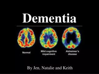
MRI in established AD Hippocampal atrophy Healthy elderly AD
Differing patterns of temporal atrophy in Alzheimer’s disease and semantic dementia C. J. Galton, MRCP(UK);, K. Patterson, PhD;, K. Graham, PhD;, M.A. Lambon-Ralph, PhD;, G. Williams, PhD;, N. Antoun, FRCR, FRCP;, B.J. Sahakian, PhD and J.R. Hodges, MD, FRCPNeurology 2001 57: 216-225
18FDG: a PET tracer analogue of glucose glucose-6-phosphate glucose Energy FDG-6-phosphate FDG blood vessel Brain cells Metabolic pathways of glucose and 18Fluorodeoxyglucose
The image Healthy brain areas need glucose and thus appear bright on the scan Damaged brain areas are not working and therefore do not pick up glucose
Alzheimer’s disease Co-registration Spatial normalisation to standard template Smoothing Statistics
Very mild Alzheimer’s-Isolated memory impairment Posterior cingulate hypometabolism
Retrosplenial cortex is the first area universally affected Nestor et al, Eur J Neurosci 2003
60 year old male, recent onset memory impairment, MMSE = 30/30
Anatomy Cingulum bundle
Regions of interest traced onto 3T volumetric MRI. FDG-PET co-registered onto MRI CMRglc calculated Normalised to cerebellum 3-compartment partial volume correction Method
Pathology Neuronal loss Amyloid deposition ACh activity loss NFT (t) Marker [18F]FDG [11C]PIB [11C]PMP In development Alzheimer’s disease and positron emission tomography