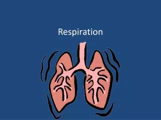
Respiration
Respiration. B&S Chapter 18. Respiration. Not just breathing in and out Or inhaling and exhaling… Respiration is the process by which oxygen is obtained from the environment and delivered to cells and carbon dioxide is transported to the outside in a reverse pathway.

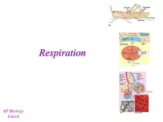
Respiration
E N D
Presentation Transcript
Respiration B&S Chapter 18
Respiration • Not just breathing in and out • Or inhaling and exhaling… • Respiration is the process by which oxygen is obtained from the environment and delivered to cells and carbon dioxide is transported to the outside in a reverse pathway
Respiration includes 3 phases: • 1. Pulmonary ventilation • 2. Diffusion • 3. Transport of gases
Pulmonary ventilation • This is the exchange of air between the atmosphere and the air sacs of the lung • Pulmonary ventilation is usually accomplished by the inspiration and expiration of breathing
The Diffusion of Gases • This includes the passage of oxygen from the air sacs into the blood and the passage of carbon dioxide out of the blood
Transport of gases • Into the circulating blood. O2 is carried to the cells and carbon dioxide is transported from the cells to the lungs
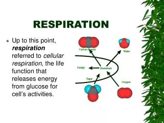
Cellular respiration • This is at the cell level… • O2 is taken into the cell and used in the breakdown of nutrients with the release of energy • CO2 is the waste product of cellular respiration
The Respiratory System • Air travels into the nostrils at 21% room air • So…of the 100% of the air in the room, we need 21% oxygen at minimum, to breathe and be 95-100% • The atmosphere we are in right now only has 21% O2 in it and this is fine for normal, breathing humans
The Respiratory System • Air travels into the nasal cavity • Moves to the pharynx (3 parts) • To the larynx • Over the Adam’s Apple (voice box) • Into the trachea • To the carina • To each left and right bronchi • To the bronchioles • To the alveoli
Air enters the nasal cavity which is divided by a septum • The septum and the walls of the nasal cavity are lined with mucous membrane • There are many blood vessels here. These vessels provide heat and moisture to the inhaled air • Cilia act as filters to dust and other debris
Mucous Membrane • The cells of this membrane secrete a large amount of fluid, up to 1 quart a day, this helps keep the nasal cavity moist • If the mucosal membrane is dry, small microscopic cracks form and this is an easy entrance for microorganisms
On the side walls of each nasal cavity are three projections called conchae The shell-like conchae greatly increase the surface area over which air must travel on its way through the nasal cavities Conchae (konk-ke)
Pharynx – throat • There are 3 parts: • Nasopharynx • Oropharynx • Laryngeal pharynx • All parts are made of muscle and help to carry food and liquids into the digestive tract
Nasopharynx • This is the top part of the pharynx, it is behind the nasal cavity
Oropharynx • This is the middle section of the pharynx and is located behind the mouth • This is the “throat” as we know it
Laryngeal Pharynx • This is the lowest portion of the pharynx • This last section opens into the larynx toward the front and into the esophagus towards the back
Larynx • A.K.A. voice box • This is located between the pharynx and the trachea • It is made of cartilage (partly thyroid cartilage), that protrudes in the front of the neck
The projection formed by the thyroid cartilage is popularly called the Adam’s Apple because it is bigger in the male than it is in the female Adam’s Apple
On both sides of the larynx… • There are folds of mucous membrane used in producing speech • These are the vocal folds or vocal cords • They are set into vibration by the flow of air from the lungs
Deep Voice • A difference in the size of the larynx is what accounts for the difference between male and female voices • Men have a lower pitch than women • The nasal cavities, sinuses, and pharynx all serve as resonating chambers for speech
The space between the vocal cords is called the glottis The little leaf shaped cartilage that covers the larynx during swallowing is the epiglottis, this protects us from aspiration Glottis & Epiglottis
Swallowing • As the larynx moves upward and forward during swallowing • The epiglottis moves downward covering the opening of the larynx • The glottis assists by closing during swallowing • Gently place your fingers over your larynx as you swallow, you will feel the movement
Trachea • Also called the windpipe • Purpose of this structure is to conduct air between the larynx and the lungs • The trachea has a framework or ringed cartilage to help keep it open, the rings are “C” shaped and are found along the entire length of the trachea
Carina • At the bottom of the trachea is the split to where the Right & Left bronchi are • This is the point to which you push the suction catheter down to, if you go any further down, your catheter will enter the Right or Left main bronchi…this is not comfortable for the pt
Bronchi • Right Bronchus: larger in diameter than the left • Extends downward in a more vertical direction • If a foreign body does get inhales in, it is likely to go to this right side first d/t the angle and gravity • Each bronchus enters the lung at a notch or depression called the hilus
Hilus • This is the place where blood vessels and nerves connect with the lung in this region • Hilus is on either right or left bronchus
Lining of the bronchus • These are lined with a special type of epithelium cells called: • Simple columnar epithelium • The cells are arranged in these columns that make it look stratified like stripes • The epithelial cells contain cilia to dust out the impurities and to create movement of fluids within the conducting tubes
Cilia in the bronchi • Help to sweep impurities toward the throat where they can be eliminated by coughing, sneezing or blowing of the nose
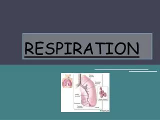
The Lungs • This is the organ in which the diffusion of gases takes place through extremely thin and delicate lung tissues • The bronchi split into right and left lobes of the lung • Right has 3 lobes • Left has 2 lobes
The bronchi further subdivide into tiny structures that resemble trees therefore, they are called bronchial tree Bronchial tree
Bronchioles • As the bronchi get smaller, they branch into bronchioles • The bronchi contain small bits of cartilage to help keep them open, there are smaller amt of cartilage the deeper you go into the bronchi. • In the bronchioles, there is NO cartilage, it now becomes smoothe muscle which is under the control of the ANS = involuntary
The alveoli • There are millions of alveoli at the end of the terminal bronchioles. They are the smallest division of the bronchiole tree, there are clusters or sacs which hold air, known as alveoli • The alveoli are thin walled and single celled and they provide easy passage for gasses entering and leaving the blood as the blood circulates through the millions of tiny capillaries that cover the alveoli
Surfactant • Certain cells in the alveolar wall produce this substance that reduces surface tension or the pull of fluids that line the alveoli….the substance is called surfactant • The surfactant prevents collapse of the alveoli and eases expansion of the lungs
How does blood travel to the alveoli • The pulmonary circuit brings blood to and from the lungs (pulmonary artery takes blood through the lung from the heart then sends it back to the left atrium via the pulmonary veins) and as blood passes through the alveoli, gas exchange takes place
Diaphragm • The lungs take up a great deal of space in the thoracic cavity • It is a large dome-shaped muscle that is attached to the body wall around the base of the rib cage • The lungs are separated from the abdominal cavity by the muscular partition known as the diaphragm • The diaphragm moves downward in the thoracic cavity when we inhale, this allows for more room to allow the lungs to expand
What covers the lungs… • A continuous double sac known as the pleura covers each lung, the pleural sac is made of serous membrane • The different areas that the pleura covers are called different things such as: • Parietal pleura is the portion of the pleura that is attached to the chest wall • Visceral pleura is the portion that is attached to the surface of the lung
Pleural Space • Between the parietal and visceral layers of the pleura is called the pleural space • This space hold the serous fluid that lubricates the 2 membranes to avoid friction when the lung expands, this allows the lungs to open bigger effortlessly
This is the region between the lungs, it contains the heart, great vessels, esophagus, trachea and lymph nodes Mediastinum
Intercostal Muscles • The diaphragm is very important in enlarging the size of the thoracic cavity to allow the lungs to inflate… • The intercostal muscles also help. They contract and lift the rib cage upward and outward • (Costochondritis)
What happens when we breathe… • Inhalation – is considered to be the “active” phase, air rushes in expanding the lungs and moving the thoracic cavity making the pressure in the pleural space drop causing air to be drawn into the lungs as by suction • Exhalation – is considered to be “passive”. The muscles of respiration relax and the lungs recoil and we breathe out carbon dioxide
When O2 enters the bronchi…. • It travels down through the bronchiole tree to the bronchioles and down to the alveoli • Here, the incoming air mixes with the existing air that’s already in the alveoli so that the gasses soon are evenly distributed • New O2 comes in and CO2 moves out in both blood and out of mouth into the air