Download
1 / 50
520 likes | 842 Vues
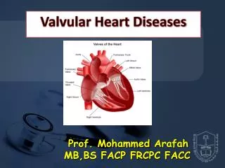
Anaesthesia For Valvular Heart Diseases. Made by: Dr. Meenal Aggarwal Moderator: Dr. Aparna. Introduction. Valvular ds : An increased burden on L or R ventricle Could be: Pressure overload ( Stenotic lesions) Volume overload ( Regurgitant lesions)

E N D
Anaesthesia For Valvular Heart Diseases Made by: Dr. MeenalAggarwal Moderator: Dr. Aparna
Valvulards: An increased burden on L or R ventricle • Could be: • Pressure overload (Stenotic lesions) • Volume overload (Regurgitant lesions) • Initially tolerated d/t compensatory mechanisms Eventually cardiac muscle dysfunction CHF ; even sudden death
Preoperative Assessment • Aim: to know • Severity of Disease • Degree of impaired myocardial contractility • Presence of assoc. organ system diseases • O/H: Symptoms: • Dyspnea, orthopnea, easy fatiguability (Impaired myocardial contractility) • Anxiety, diaphoresis, resting tachycardia (Compensatory increase in sympathetic activity) • Angina (d/t assoc CAD, or inc. myocardial O2 demand)
Drug therapy: • Beta Blockers • Digitalis • ACE inhibitors • Vasodilators • Diuretics • Ionotropes • Antiarrhythmic drugs • Control HR (AS & MS: Allows diastolic filling) • Control BP and so dec. afterload (AR, MR) • Control of CHF
O/E: Signs: • Inspection: Raised JVP • Auscultation: Basilar chest rales, S3, Murmurs • Murmurs: D/t turbulent flow across the defective valve • Note: character, location, intensity, direction of radiation • Systolic murmurs: AS, PS or MR,TR • Diastolic murmurs: MS, TS or AR, PR • Dysrhythmias: AF (esp Mitral valve ds.) i.e. with enlarged Lt atria
Lab Investigations: • CXR: • Size & shape of heart & great vessels • Pulmonary markings • Enlarged LA (Elevated Lt main bronchus, calcified valve) • ECG: • Lt or Rt axis deviation (Lt or Rt ventricle hypertrophy) • P mitrale (Broad notched P wave in Mitral valve ds.) • Dysrhythmias • Conduction abnormalities • Evidence or active ischemia or previous MI
Echo with doppler: • Evaluating significance of murmurs • Detection of antomical defects (Hypertrophy, chamber size, valve area) • Functional defects (Transvalvular pressure gradient, magnitude of valvular regurgitation) • Cardiac Catheterisation: Solves discrepancies b/w clinical and echo findings • Presence & severity of stenosis or regurgitation • Intracardiac shunting • CAD
Transvalvular pressure gradient (TVPG) (Severe MS when > 10mmHg, Severe AS when > 50 mm Hg) • Pulmonary artery pressures (Pulmn HT) • Assessment of Prosthetic Valve function: • Dysfunction (Change in intensity/ quality of clicks, new or change in characteristics of murmurs) • Tranthoracic Echo: To assess ring stability and leaflet motion • Transesophageal Echo: Better resolution • MRI: For prosthetic valve regurg, paravalvular leak • Cardiac Catheterisation: For TVPG, Effective valve area
Complications of prosthetic valves: • Risk of thromboembolism (Anticoagulation) • Subclinical intravascular hemolysis • Risk of endocarditis (AB) • Management of anti coagulation: • Can be continued in minor surgery with min blood loss • For major surgery (Stop warfarin 3-5 days preop, UF heparin or LMWH started & continued upto day/ day before of surgery, restarted post op) • Avoid elective surgery with in 1 month after an acute thromboembolic episode
In pregnancy (TE prophylaxis to continue, S/C LMWH given + low dose aspirin) • Prophylaxis of Bacterial endocarditis: • Infection likely from frequent exposure to bacteremia • Weigh Risk to benefit ratio (AB resistance) • Prophylaxis given to following pts: • Prosthetic material for cardiac valve repair • Previous IE • CHD: Unrepaired CHD, Completely repaired with prosthetic material (during 1st 6 months after procedure), Repaired defects with residual defect) • Cardiac transplant pt who develop valvulopathy
AB prophylaxis not required for GU or GIT procedure • Required for skin incision/ Biopsy or Resp tract invasive procedure • For dental procedures (manipulation of gingiva, Mucosa)
MITRAL STENOSIS: • Most common cause RHD • Primarily affects females • Diffuse thickening of mitral leaflets & subvalvular apparatus, Calcification • Gradual progression (over 20-30 yrs) • Other causes: Carcinoid syndrome, LA myxoma, Severe mitral annular calcification, RA, thrombus formation, SLE, congenital
Decreased mitral valve orifice • Mechanical obstruction to LV diastolic filling • Dec LV volume • Inc LA volume & pressure • Dec S.V. • Inc Pulmn Venous Pressure • RV Hypertrophy & failure Overt Pulmn Edema • Pathophysiology of Mitral Stenosis
Diagnosis: • Clinical signs: opening snap (in early diastole), rumbling diastolic heart murmur • Venous thrombosis (stasis, decreased activity) • CXR: -LA enlargement (straightening of left heart border, elevation of left main stem bronchus, double density of LA) • -Mitral calcification • -Evidence of pulmn congestion • ECG: Broad notched P wave (P mitrale), AF
Echo: (Anatomical details: Leaflet thickening, calcification, changes in mobility, chamber dimension, thrombus) • Severity assessed by: • - Mitral valve area, TVPG • Also for Pulmn HT, Ventricular function
Treatment: • Mild MS: Diuretics • In AF: Beta blockers, Ca #, Digitalis (H.R. control) • Anticoagulants (Warfarin to get INR of 2.5 to 3) • Surgical correction: • Percutaneousvalvotomy • Valve reconstruction • Valve replacement, surgical commisurotomy
Management of Anaesthesia: • Avoid tachycardia (prevents filling) • Avoid decrease in SVR (use vasopressors which avoid Tachycardia) • Do no permit volume overload (can ppt CHF) • Prevent hypercarbia & hypoxemia, lung hyperinflation (Worsen Pulmn HT) • If RVF : Requires ionotropic support & pulmonary vasodilators • Premedication: decrease anxiety (watch for resp depression), Continue drugs for HR control, Treat diuretic induced hypoK+
Anticoagulant therapy (acc to minor or major procedure), coagulation tests for regional anaesthesia • Induction: I/V agents (except ketamine), MR (which doesn’t Inc HR or Dec BP d/t histamine release) • Maintenance: Min effect on HR, SVR & PVR, contractility (N2O+ opioid+ Low conc Volatile agents) • Reversal achieved slowly (to avoid tachycardia d/t glyco/atropine) • Prevent light plane of anaesthesia (symp stimulation) • Pulmonary vasodilator may be required • Careful fluid replacement intraop (risk of Pulmn edema)
Monitoring: In asymptomatic (routine) • Symptomatic/ major surgery (Intraarterial pressure monitoring, Pulmonary artery pressure, LA pressure: at higher risk of rupture of pulmn A so done carefully and less frquently, TEE) • Post operative management: • Prevent fluid overload • Manage pain (to prevent tachycardia, hypoventilation so hypoxia), neuraxialopioids • May require mechanical ventilation (thoracic surgery)
MITRAL REGURGITATION: • In RHD, usually assoc with MS • Other causes: Papillary muscle dysfxn, mitral annular dilatation, rupture of chordaetendinae, endocarditis, MVP, Congenital • Pathophysiology: • Regurgitation into LA LA volume overload Dec LV stroke volume LA enlargement & AF • Pulmn congestion
Regurgitant fraction depends on: • Size of valve orifice • Heart rate • Pressure gradient across MV (SVR) • When MR develops gradually: LV becomes more compliant • When acute MR: No compensation, sudden sever Dec in S.V. l/t cardiogenic shock, with pulmn congestion • When MR+ MS : both volume and pressure overload • Diagnosis: • O/E: holosystolic apical murmur, radiation to axilla • CXR: Cardiomegaly (LA & LV hypertrophy)
Diagnosis cont… • ECG: Lt axis deviation • Echo: Confirms MR, Anatomy (LA size, LV wall thickness, cavity dimension), S.V., LA appendage for thrombus • Doppler: Severity assessment (Calculation of regurgitant volume and fraction), area of regurgitant jet • Pulmn A. Occ. Pressure: Shows a ‘V’ wave in the waveform signifies regurgitation • Cardiac catheterisation: If surgery planned or severity doubtful • Coronary angiography: In elderly patients
Treatment: • Surgical: • Mitral valve repair (preferred as apparatus preserved) • Mitral valve replacement • Survival increased by surgery of performed before LVEF < 60%, or before End systolic LV dimension >= 45mm • Patients who do not improve with surgery: • * LVEF < 30% * LV end systolic dimension > 55mm • Medical : • Vasodilators (Acute MR) • Beta #, ACE inhibitors • Biventricular pacing
Management of Anaesthesia: • Prevent events which Dec C.O. • Maintain N to slightly higher H.R. • Vasodilators to decafterload • Ionotropes to improve LV contraction • Induction: • I/V agent used • MR (pancuronium beneficial- raises HR) • Maintainence: • Inhalational agents (Dec rise in BP & SVR caused by surgical stimulation) iso, des, sevo
Opioids (when severely compromised myocardial function) • Mechanical ventilation (allow venous return) • Maintain I/V volume • Monitoring: • Asymptomatic / minor surgery (no invasive monitoring) • Severe MR (Pulmn A. Catherisation V wave)
MITRAL VALVE PROLAPSE: • Prolapsed one/ both mitral leaflets into LA during systole • M.C. form of valvulards. (young women) • With or Without MR • Causes: Marfan’s, RHD, Myocarditis, thyrotoxicosis, SLE • Diagnosis: • Usually benign, but can l/t IE, cerebral embolisation, Severe MR, Severe dysrrhythmias, sudden death • C/F: Palpitation, anxiety, orthostatic symptoms, dysnea, fatigue, atypical chest pain
Echo: valve prolapse of 2mm or more above mitral annulus • With/ without leaflet thickening (elderly/connective ts. ds) • Functional form (mild bowing) • Management of Anaesthesia: • Influenced by degree of MR • Basis: Larger LV will have lesser prolapse • Inc sympathetic activity • Dec SVR • Upright posture • hypovolemia Increase MR
Inc LV vol will Dec MVP (HTN/ Vasoconst, drug induced myocardial depression, volume resuscitation) • Preoperative Evaluation: • Differentiate functional MVP from significant MR • Usually< 45 y, female • Beta blocker for arrhythmias (continued) • If H/O Transient neurological event with sinus rhythm, no atrial thrombi (pt usually on aspirin 81-325mg/d) • Pt with AF &/or with atrial thrombi or previous stroke (usually on warfarin) • ECG changes (PVC’s, QT prolongation) no implication
Pt may have systolic clicks, murmur even without symptoms (no need of cardio consultation) • In older men (MVP can present with CHF) pt on diuretics, ACE inh • Anaesthesia technique: • When LV function normal, tolerates both GA & regional • Induction: • I/V agent (assess need to avoid dec in SVR) • Etomidate (min Myocardial depression) • Ketamine not to be used (Enhances LV emptying so inc MR) • Maintenance: • Minimize sympathetic nervous system activity d/t surgical stimuli
Volatile anaesthetics with N2O +/- Opioids • Low dose: 0.5 MAC (iso, des, sevo) in significant MR • Any MR (keep in mind vagolytic/ histamine induced effects) • Unexpected ventricular arrhythmias can occur intra op (Beta blocker or lignocaine) • Proper fluid balance • Vasopressors may be required • Avoid controlled hypertension technique (increases MVP) • Monitoring: • Routine • Significant MR/ LV dysfunction (Pulmn A. catheter)
AORTIC STENOSIS: • Degeneration & calcification of leaflets (ageing), then stenosis • Causes : Elderly, Bicuspid Aortic Valve • N valve area: 2.5-3.5 cm2 • Almost always assoc with some AR
Angina may occur despite absence of CAD (Inc myocardial demand, dec supply) • Syncope (fall in SVR can’t be compensated by inc C.O.) • Diagnosis: • C/F: angina, syncope, dyspnea on exertion • O/E: Systolic murmur best heard in aortic area (be careful as mostly patients undiagnosed) • CXR: Prominent ascending aorta • ECG: LV hypertrophy • Echo with doppler: Bileaflet aortic valve, thickening/ calcification of aortic valve, decreased mobility, LV hypertrophy
Echo cont… • Valve area, TVPG • Cardiac Catheterisation • Coronary Angiography • Exercise stress testing for Asymptomatic patients • Treatment: • Asymptomatic: Continue medical therapy (delay Surgery untill s/s appear) • Aortic Valve replacement • Coronary revascularisation (if co-existant CAD) • Percutaneous aortic balloon valvuloplasty
Management of Anaesthesia: • Maintain N sinus rhythm • Avoid bradycardia/ tachycardia • Avoid hypotension (if occurs aggressive Tt required) • Optimise I/V fluid volume • CPR is generally ineffective in AS (Not enough CO generated) • Induction: • GA preferred (regional causes Hypotension) • I/V agents used (ones which do not dec SVR) • If LV function compromised opioid induction
Maintenance: • Avoid drugs which suppress S.A.node (if occurs give atropine/ glyco/ ephedrine) • If persistent tachycardia use esmolol • In supravent. tachycardiascardioversion to be done • Chanced of VT present (Lidocaine & defib) • If LV dyfxn (avoid drugs depressing myocardial contractility) • NM blocker with min hemodynamic effects • I/V fluid vol to be maintained • Monitoring: • ECG, Intraarterialcath, P.A. cath, TEE
AORTIC REGURGITATION: • Causes: IE, RF, Bicuspid aortic valve, ds of root of aorta
Magnitude of regurgitation depends on: • Time available for regurgitation (H.R. dependent) • Pressure gradient across the valve (SVR dependent) • Diagnosis: • C/F: Dysnea, orthopnea, fatigue, coronary ischemia • O/E: Diastolic murmur (Lt sternal border), bounding pulses, wide pulse pressure, Austin Flint murmur (low pitched diastolic murmur) • CXR & ECG: LV enlargement & hypertrophy • Echo: LVEF & ESV, Severity of regurgitation (on doppler) • Cardiac cath & MRI
Treatment: • Surgical: • Replacement (even in asymptomatic) Immediate surgery in acute AR (as l/t sudden heart failure) • Ross procedure (Pulmonic valve autograft) • Valve reconstruction • Medical: • Vasodilators (Nitroprusside) • Ionotropes (Dobutamine) • Long term Nifedipine/ Hydralazine
Management of Anaesthesia: • Avoid bradycardia (HR above 80/min), use atropine • Avoid inc in SVR • Minimize myocardial depression • If LV failure (vasodilators and ionotropes) • GA chosen • Induction: I/V agent which doesn’t inc SVR or dec HR • Maintenance: N2O + volatile agent &/or opioid • Iso, Des, Sevo good (inc HR, dec SVR, min myo depression) • If severe LV dysfunction high dose opioid (caution: bradycardia) • NM blocker: Pancuronium useful, modest tachycardia
Monitoring: • Minor surgery with asymptomatic ds. (routine) • Severe AR: • Pulmonary A catheter • TEE • Useful for guiding I/V volume replacement, detecting myocardial depression, measuring response to vasodilators
TRICUSPID REGURGITATION: • Usually functional (d/t RV enlargement or Pulmn HT) • IE, Carcinoid, RHD, Ebstein anomaly • Mild TR in highly trained athletes • Pathophysiology: • Regurgitation through TV RA vol Overload (but minimal rise in RA pressure) • O/E: Raised JVP, Hepatomegaly, ascites, edema • Tt: Tt the cause (improve lung fxn, relieve LV failure, dec PHT) • Surgery (rarely for TR alone), Tricuspid annuloplasty/ valvuloplasty/ replacement
Management of Anaesthesia: • Keep CVP to high Normal • IPPV may decrease venous return • Avoid hypoxemia & hypercarbia (to prevent inc Pulmn A. pressure) • N2O: weak Pulmn A. vasoconst (may inc TR) • Intra op measurement of RA pressure to guide fluid therapy • Very high LA pressure can l/t R L shunt (patent foramen ovale)
TRICUSPID STENOSIS: • M.C.cause: RHD (coexiztant TR, Mitral n aortic valve ds) • Inc RA pressure & pressure gradient b/w RA & RV • PULMONARY REGURGITATION: • Secondary to Pulmn HT • Rarely symptomatic • PULMONARY STENOSIS: • Usually congenital (detected and treated in early childhood) • C/F: Syncope, angina, RV Failure • Tt: surgical valvotomy