Download
1 / 73
740 likes | 1.04k Vues
Explore the world of fungi, from their biology to their role in skin disorders. Learn about dermatophytons, yeasts, Candida, molds, and the pathogenic nature of some fungi. Understand their classification, reproduction, and impact on human health. Discover the diversity of fungi and their interactions with living organisms in this comprehensive guide.

E N D
PROLOGUE: A BIOLOGICAL GUIDE • For a long time fungi were regarded as a primitive branch of the vegetable kingdom. Nowadays they are viewed as a separate group, alongside plants, animals, protists and prokaryote organisms. • Fungi are everywhere, occurring both in the sea, in fresh water and on land. There are some 100,000 known species. They constitute an important link in the organic chains of nature by converting dead and decaying matter into new raw materials for plants.
FUNGI AND YEASTS • In principle, the term 'fungi' is the name given to a large group of organisms with biological characteristics such as the presence of a cell membrane, a cell wall and a eukaryotic nucleus, and the absence of chlorophyll (the green colouring matter of plants). This means that the yeasts also form part of the fungi group. Strictly speaking, therefore, it is incorrect to speak of 'fungi and yeasts'. For the practicalities of daily life, however, a pragmatic subdivision has been devised in which yeasts are called 'yeasts' and nonyeasts are referred to as 'fungi'. Furthermore, the term 'fungus' usually has a negative undertone, whereas yeasts are viewed in a somewhat more positive light. Bakers' yeast is an essential ingredient, but bread mould -- the fungus that often grows on stale bread -- indicates decay. • Less than 100 of the approximately 100,000 species of fungus are pathogenic for human beings.
FROM SAPROPHYTE TO PARASITE • The absence of chlorophyll (the green pigment in plants) is central to the way in which fungi grow and live, for it precludes CO2-assimilation and photosynthesis. Fungi are therefore dependent upon an exogenous source of organic matter for their own metabolism. Usually this is dead or decaying organic waste. • As long as the growth of fungi is not achieved at the expense of living tissue, we call them saprophytes. Otherwise, i.e. when they attack living plants or animals, we speak of a parasitic lifestyle. And that can cause a fungus to become pathogenic. • Nevertheless, there are more aspects involved than the properties and lifestyle of the fungus alone. A fungus almost never infects a host without reason. There has to be a favorable climate, and that is largely determined by the host himself. The properties of the host, for example the environment of the skin and its defense mechanisms, are extremely important.
FORM AND REPRODUCTION • Fungal cells have a cell wall and a true nucleus, which means that they are classified as one of the eukaryotes. Unlike higher animals and plants, the fungi have only a single set of genetic material: they are haploid. • From the morphological viewpoint the fungi constitute a heterogeneous group of organisms. On the one hand, there are the multicellular forms which grow as ramified threads or filaments, called hyphae, with both sexual and asexual reproduction. On the other hand there are the single-celled organisms, the yeasts, whose principal method of reproduction is asexual. Some yeasts also form ramified but nonmulticellular structures which are designated by the term 'pseudohyphae'. • Sporulation is an important property of fungi. In that form they can easily survive. When a flourishing period is followed by an interval of adverse conditions, the spores can then be disseminated; they subsequently remain in a state of rest until a favorable environment is restored. This is why fungi can be obstinate and difficult to control.
CLASSIFICATION OF THE FUNGI • A definitive classification is not possible until the perfect stage has been identified and described. The classification is arranged in six groups (see table), three of which are relevant to disorders in human beings. • The last group, Fungi Imperfecti, is a group with no identified sexual reproduction and whose perfect stage is therefore unknown. • The actinomycetes are gram-positive bacteria which cause, inter alia, pseudomycoses.
Etiology of dermatomycoses • Dermatophytons of 3 genera: Trichophyton, Microsporum and Epidermophyton • Keratophytonsof some yeast species: Pityrosporum ovale, Pityrosporum orbiculare,Malassezia furfur. • Candida genus (Candida albicans). • Pseudofungi (Corynebacterium minutissimum and Actinomyces israelii. • Moulds (Scopulariopsis, Aspergillus, Penicillium, etc.)
FUNGI AS PATHOGENIC ORGANISMS • Of all the known species of fungus, only a few are pathogenic. Furthermore, the presence of such a fungus in or on the human body does not automatically mean that there is mention of pathology. The presence of Candida spp., for example, can be demonstrated in human faeces without there being any question of an infection. Fungi are therefore not inevitably pathogenic. The truth of this statement is underlined by the increasing number of immuno-compromised patients. AIDS, transplantations and chemotherapy can lead to reduced immune system function. Precisely at times such as these, fungi seize their opportunity and launch an attack. At first sight the result may appear to be a harmless case of thrush, but it may also be a systemic infection which is difficult to treat, such as a disseminated Aspergillus infection, sometimes with a fatal outcome. • Disorders caused by fungi are called mycoses. They are usually classified according to the site where they appear. There is a large group of disorders which affect the skin. Many fungi, such as the dermatophytes, go no deeper than the horny layer of the skin: these are the superficial infections. When fungi are also demonstrated deeper in the tissues, these are referred to as subcutaneous infections. If organic systems or organs are affected, they are called systemic infections. It will be obvious that the latter group is the most life-threatening. In terms of frequency, however, systemic fungal infections are the rarest.
DERMATOMYCOSES - DEFINITION AND CHARACTERISTICS • Dermatomycoses are infections of the skin, hair or nails by fungi. The principal causative agents are dermatophytes, which are subdivided into three groups (genera): Microsporum spp., Trichophyton spp. and Epidermophyton floccosum. The three genera are distinguished by the form of the spores, or macroconidia. • Trichophyton: thin-walled, smooth, four to six septa • Microsporum: thick-walled, with projections five to more septa • Epidermophyton: thick-walled, pear to oval shaped four or fewer septa • Besides the dermatophytes, yeasts are also capable of causing skin disorders. The most frequent agents in this case are Candida spp. and Pityrosporum. • Infections are increasingly being caused by species of fungus which are classified neither as yeasts nor dermatophytes – moulds. An example of this is Scopulariopsis brevicaulis, which can occur in nails.
DERMATOPHYTES - DEVELOPMENT OF A DERMATOPHYTOSIS • As already mentioned in the introduction, fungal infections do not occur without reason. • As dermatophytes are not commensals, a prerequisite for the development of an infection is exposure to the fungus. This is possible, for example, by direct contact with infected persons or animals, but it is more often a question of contact with fungal spores. These spores are contained in epithelial (skin) elements of infected persons everywhere in our environment. The floors of communal shower stalls and changing rooms are major sources of infection. For the development of an infection, however, more is needed than contact alone. • Dermatophytes prefer warm, moist conditions. This is why a dry, intact skin constitutes a virtually impenetrable barrier. But the chance of infection is encouraged by everything that has an adverse influence on the situation.
DERMATOPHYTES – THEIR HOST • Dermatophytes do not have an exclusive preference for human beings. Some of the infections in humans even originate in (domestic) animals. • On the basis of the original host, a distinction is made between anthropophilic, zoophilic and geophilic dermatophytes. This distinction is very important, chiefly because in the event of infection of human beings by zoophilic dermatophytes, the source of the infection (the animal) must be co-treated. • In the case of geophilic infections one can try to avoid further contact with the source. • A point worthy of mention is that zoophilic dermatophytes in human beings frequently evoke a more intense inflammatory reaction than an infection by anthropophilic species. The latter have adapted themselves better to life in the human epidermis and are regarded to a lesser degree as invasive organisms which have to be opposed.
DERMATOPHYTES - PREFERRED SITES OF INFECTION • Most dermatophytes have been found to have a preference for certain situs. A preference for growth in and around the hair, in the horny layer of skin, in the moist, warm folds of the skin, or just under the nails. Trichophyton species have been found to have the greatest adaptability, or perhaps they are merely the least fastidious. They are capable of causing tinea capitis, corporis, barbae, pedis, plantaris and tinea unguium. • Epidermophyton floccosum occurs principally in the large flexure lines and around the foot. Microsporum chiefly attacks the scalp and glabrous skin. It rarely occurs in flexure lines or the nails. Furthermore, the preferred sites of infection of dermatophytes are, to a certain extent, also determined by the situs where the skin comes into contact with the fungal spores.
KERATINOPHILIA • A major characteristic of dermatophytes is their keratinophilia. They only grow in the dead, horny layer of the skin. They do not reach deeper tissue or deeper parts of the skin. Dermatophytes can only be found in the epidermis, hair shaft, nail plate and nail bed. However, this does not mean that no changes occur in the deeper layers of skin. After all, the clinical picture that emerges is caused by the inflammatory reaction which occurs deeper in the dermis. • Now, it seems fairly logical to assume that keratinophilic fungi, such as dermatophytes, do not grow deeper into the skin because there is no keratin present in the deeper layers. However, it is open to question whether this keratinophilia also means that these fungi are keratin-dependent. There are indications that it is not the absence of keratin but rather the presence of serum factors that inhibits the growth of dermatophytes deeper into the skin. It is also not known whether keratin is converted by an enzyme (a keratinase) of the dermatophyte itself. It is presumed that mechanical factors -- by pressure from the hyphae themselves -- also damage the keratin.
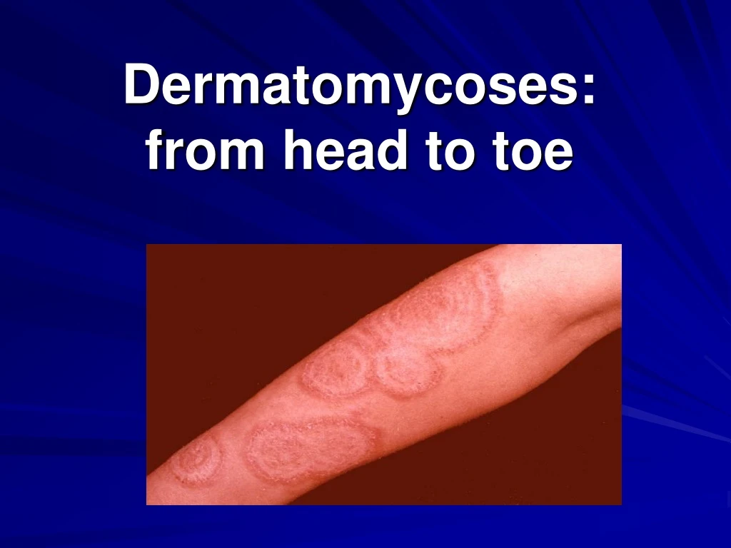
DERMATOPHYTES AND THE SKIN • The classical picture of a dermatophyte infection of the skin is a round patch with peripheral activity: the fungus spreads peripherally. This process is attended by the formation of waste matter and toxins which function as a mediator in inflammatory reactions. The inflammation clears towards the centre and there is a progressive decline in the number of hyphae which are found. The conditions for the growth of fungi apparently become less favourable. Fewer hyphae does not however mean that there are also fewer fungi. Obviously, the central hyphae are older and spores have been formed within them. In this case the hyphae assume the form of a string of beads and then fragment. These elements are much more difficult to find by microscopy than hyphae. • It is still not clear why dermatophytes grow almost exclusively towards the periphery. In some way or another, perhaps immunological, the central portion offers more resistance or is less attractive to the dermatophyte than skin which has not yet been infected.
DERMATOPHYTES AND THE NAILS • A dermatophyte infection of the nail often starts at the free margin of the nail or the lateral nail fold. Where the feet are concerned, these are precisely the places where pressure is exerted by footwear and where damage can readily occur. From there the dermatophyte grows into the nail plate to the base of the nail, as a result of which the nail plate can become thickened, friable and even totally destroyed, forced up by the underlying hyperkeratinization. • One result of the infection of this particular nail is that nail growth is inhibited. And that growth was already fairly limited: dermatophyte infections have a predilection for nails which have a poor growth rate due to other reasons. And since nail growth is essential for a cure, obviously the healing process is prolonged. During treatment and the healing process, the dermatophyte retraces its steps as it were: from the base to the free margin of the nail. Besides the difference in direction, however, there is also an important difference in the rate of activity, for the gradual replacement of the infected nail is now determined by the (much slower) growth rate of the nail and not by the (high) growth rate of the dermatophyte. The development of an onychomycosis therefore progresses much faster than the corresponding healing process.
DERMATOPHYTES AND THE HAIR • A number of dermatophytes can penetrate into the hair follicle and some of them can even invade the hair. The process by which dermatophytes grow, which helps to explain the clinical picture, can be examined under the microscope. • Endothrix: In this form of growth the fungus develops from the stratum corneum into the follicle. The hyphae subsequently develop in the hair shaft and attack the entire structure of the hair. The fungus grows further into the radix pili, or root of hair, as a result of which the hairs are filled, as it were, with mycelial threads and spores ( see illustration a ). Owing to the growth of the hair itself, the swollen and spore-filled portions of the hair shafts reach the surface of the skin, where they break off. There is therefore no question of stubble. This form of growth, with the presence of spores in the hair, is known as endothrix growth and occurs, inter alia, in infections by Trichophyton violaceum.
DERMATOPHYTES AND THE HAIR • Ectothrix: The second form of growth is called ectothrix growth. In this case, the hyphae tend to grow primarily around the hairs, thereby producing a veritable sheath of spores. The size of the spores varies quite considerably. An example of a fungus with small-spored ectothrix growth is Microsporum canis, while Trichophyton mentagrophytes is large-spored. Ectothrix growth results in the hairs breaking off a few millimetres above the surface of the skin; this is because the basic structure is not so severely infected. This gives the skin a stubbled appearance.
DERMATOPHYTES AND THE HAIR • In favus, caused by Trichophyton schoenleinii, there is no sporulation in the hairs and limited formation of mycelium. Consequently, in this infection hairs are able to grow to their normal length. • Not all dermatophytes are capable of growing into the hair follicle: Epidermophyton floccosum is an example of such a dermatophyte. • Microsporum canis, on the other hand, has no difficulty whatever in invading the follicle.
Summary of the different forms of growth of dermatophytes in hair
YEASTS • The two major species of yeast capable of causing skin infections are Candida albicans and Pityrosporum ovale. • The most striking property of these yeasts is that they are commonly part of the normal flora. We are therefore dealing with real opportunists. This also means that the way in which an infection develops is quite different to an invasion by dermatophytes. • As mentioned earlier, with dermatophytes some form of exposure to the fungus and its spores is necessary before an infection can develop. In the case of yeast infections, however, particular importance attaches to predisposing factors.
CANDIDA • There are about 100 known species of the Candida genus. Not all Candida species are present in human beings as commensals or pathogens, not by any manner of means. It is only in exceptional cases that another yeast than Candida albicans plays a role. C. albicans is normally present as a commensal in the mouth, the gastrointestinal tract and the vagina. • When is there mention of an infection (e.g. thrush or vaginal candidosis) and what changes are involved? In an infection there is a multiplication of the number of yeast cells: far more than when Candida is merely present as a commensal. Furthermore, commensal Candida is only present in yeast form, whereas in infections (pseudo)hyphae are also found (see image 1). And in an infection there are also signs of pathology, for example inflammation. • There are various predisposing factors which are conducive to the transition from commensal to pathogenic. A moist skin, a high pH and the presence of sugars and certain amino acids create a favourable climate for Candida. • In the host, reduced cellular immunity is a major predisposing factor. Diabetes mellitus is also frequently implicated in Candida infections. However, it is open to question whether a properly adjusted patient runs a higher risk of candidosis. A good example of the effect of sacchariferous substances is found, inter alia, among bakers and workers in the confectionery industry. Candida paronychia is an occupational disease in these people. A damaged skin is another important factor. Macerated skin, as in moist flexure lines, is a preferred site for Candida.
PITYROSPORUM • It is a lipophilic yeast which only grows when oil, glycerin or glyceryl monostearate is added to the culture medium. Pityrosporum is chiefly found as a commensal on areas of the skin containing a relatively large number of sebaceous glands. The yeast can assume various morphological forms. Until quite recently this led to the assumption that Pityrosporum ovale, Pityrosporum orbiculare and Malassezia were three different organisms. Now, however, it is known that they are different forms of growth of one and the same yeast: P. ovale. In this scenario Pityrosporum is the yeast form while Malassezia furfur, formerly always described as the causative agent of pityriasis versicolor, is the mycelial form. • In pityriasis versicolor, just as in candidosis, there are several conductive factors. The principal one is the climate. In tropical regions we see incidences of 40 to 50%, as opposed to temperate climates where not even one per cent is achieved. Furthermore, the infection usually develops in the summer months; the use of sunscreens is said to be a conducive factor. • It has recently been established that P. ovale also plays a role in seborrhoeic dermatitis. Patients with this infection have a high concentration of P. ovale on the skin, as a result of which inflammatory reactions occur. Dandruff is regarded as a form of seborrhoeic dermatitis.
DERMATOMYCOSES - CLASSIFICATION • Dermatomycoses can be classified in various ways. The simplest of all would seem to be a systematic arrangement on the basis of the causative agents. In a classification of this nature, the disorders are designated by reference to the individual genus: trichophytosis, epidermophytosis, microsporosis, candidosis (candidiasis) and pityrosporosis. • A classification focused more on the epidemiology and method of dissemination is one that is based on the original host of the various fungi. This produces such terms as anthropophilic, zoophilic and possibly geophilic agents: fungi which therefore have human beings, an animal or the soil as their primary habitat. • Closer to actual practice is a classification on the basis of the clinical picture, designated, for example, by the severity of the inflammation: mild, moderate or severe. The most widely used classification is largely based on the site of the clinical picture. As mentioned earlier, the largest group of dermatomycoses consists of disorders which are caused by dermatophytes. They are designated by the name 'tinea'. A distinction is made between the tinea group (dermatophyte infections), which is subdivided according to the site of the infection, and a group of yeast infections, in which the subclassification is on the basis of the causative agent (see table).
TINEA CAPITIS In tinea capitis, also called ringworm of the scalp, the lesions are typically ring-shaped and the skin and hair are infected. The hairs break off and leave bald patches. Four subgroups of tinea capitis can be distinguished: • - Microsporosis • - Trichophytosis (herpes tonsurans) • - Favus • - Kerion
MICROSPOROSIS • Microsporosis is an infection caused by Microsporum spp. which occurs most frequently in school-going children. It is transmitted from one child to another, but sometimes it can also be a family infection. In the event of a recurrence, it is therefore of the utmost importance to localize the source of the infection -- certainly not forgetting the family pets. The commonest Microsporum species are M. canis and M. audouinii. • Clinical picture: The disorder begins as a small plaque with thin grey scaling. There are short, broken hairs which are only a few millimetres long, producing on palpation the effect of a brush with short, stiff bristles. Sometimes there are severe inflammatory symptoms, such as erythema and swelling. The signs depend primarily on the pathogenic organism. M. canis, for example, being a zoophilic fungus, produces more severe inflammation than M. audouinii. Furthermore, there may be a concomitant tinea corporis. The plaque becomes larger within a few weeks. New lesions appear and may become confluent. After relatively rapid growth at the onset of the disorder the expansion generally comes to a halt. After puberty a spontaneous regression occurs without scars or lesions.
TRICHOPHYTOSIS • Children are especially prone to attack by trichophytosis or herpes tonsurans, but it can also infect adults. Trichophytosis is an exotic disease from Turkey and Morocco and seldom occurs in more temperate climates. The major causative agents of herpes tonsurans are Trichophyton tonsurans and T. violaceum. • Clinical picture: Clinical findings usually comprise a large number of small grey patches. In most cases erythema is absent or only mild. The patches are characterized by partial hair loss. The hairs are broken off on the surface of the skin and are only visible as a small stub. They may be found in coexistence with long, healthy hairs. The lesions are covered with a layer of grey scales and may become confluent, thereby causing an irregular pattern. As in microsporosis, spontaneous healing can occur after puberty.
FAVUS • Favus is caused by Trichophyton schoenleinii and is most frequently found in subtropical regions. In more temperature climates the disorder has practically been eliminated thanks to griseofulvin, which made effective treatment possible. • Clinical picture: Favus is a disorder with a unique clinical picture. It begins as a red scaling patch on the scalp which develops until it covers an area several centimetres in diameter. The next stage is the formation of scutula: yellow, cup-shaped crusts with a diameter of one to two centimetres.A salient feature is that the hairs are not broken off and the length of some of them is quite normal. They are nevertheless infected: the hairs lose their gloss and are arranged on the scalp in irregular tufts. As the patch increases in size, total and irreversible hair loss occurs in the central region. The 'mouse smell' is mentioned in all textbooks. The impetiginous form of favus is characterized by moist crusts with underlying accumulations of pus. A secondary coccal infection frequently occurs and makes the differential diagnosis with impetigo more difficult. There is no tendency to healing after puberty as in microsporosis.
KERION • Kerion occurs at all ages. The causative agents of this disorder are Trichophyton verrucosum and T. gypseum. • Kerion begins as an erythematous annular patch which gradually elevates itself above the surrounding skin. It is clearly circumscribed, while the slightly nodular surface is covered with pustules ( see image 3 ). Pus is released when pressure is applied. It is associated with occasional pain. A kerion infection is not restricted to the scalp. Infections of this nature are also possible in the beard area. • If left untreated, the condition will persist for several weeks or months. Then the symptoms will gradually diminish. An atrophic scar may remain after healing, while the sustained hair loss will not be fully replaced. However, superinfections can seriously complicate this relatively benign process.
TINEA BARBAE • Tinea barbae is quite often seen in dairy farmers, who become infected through contact with cows while milking. Tinea barbae is usually caused by T. mentagrophytes or T. verrucosum. • Clinical picture: Tinea barbae is also called trichophyte sycosis. In principle, tinea barbae and tinea capitis are one and the same infection. The agents are dermatophytes which attack the hair and skin, with severe inflammatory plaques possibly attended by scaling and incrustation. Kerion also occurs in association with tinea barbae. The infection can last for months and there is a real risk of bacterial superinfection.
TINEA CORPORIS • Every dermatophyte can be the causative agent of tinea corporis, in other words Microsporum spp., Trichophyton spp. and Epidermophyton floccosum. The nature of the dermatophyte may help to indicate the source. Zoophilic fungi (for example, M. Canis or T. mentagrophytes) may have been contracted from an infected pet animal. • Clinical picture: Tinea corporis, which was formerly also called herpes circinatus, is a tinea or ringworm disorder of glabrous skin. The general characteristics are annular scaling patches and a slowly expanding edge with inflammation which is frequently somewhat elevated. The lesions are clearly circumscribed and vesicles may also occur. The patient may also complain of itch and a burning sensation. The lesion spreads peripherally and tends to heal in the centre. One or several weeks after infection with dermatophyte spores, the visible lesion appears. After several months, depending in part on the species of fungus involved, spontaneous healing can occur. Chronic infections are also possible, however, and T. rubrum infections are notoriously obstinate.


![[READ DOWNLOAD] From Head to Toe](https://cdn7.slideserve.com/12541761/slide1-dt.jpg)