Download
1 / 75
840 likes | 1.58k Vues
Chapter 1 Developmental disturbances of teeth Chapter 2 Dental caries. Dental Caries نخر الأسنان : - most common of all diseases & - major cause of loss of teeth. - Is a progressive irreversible bacterial damage to teeth exposed to the saliva &

E N D
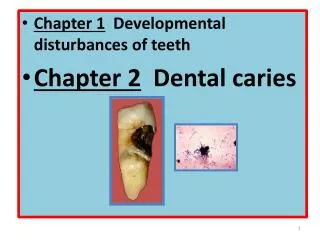
Chapter 1 Developmental disturbances of teeth • Chapter 2 Dental caries
Dental Caries نخر الأسنان : - most common of all diseases & - major cause of loss of teeth. - Is a progressive irreversible bacterial damage to teeth exposed to the saliva & initiated by acid production in bacterial plaque. Etiology of dental caries : Develops only in the presence of several interacting variables such as : 1- Cariogenic bacteria 2- Bacterial plaque. 3- Sugar. 4- Susceptibility of teeth to caries.
1) Cariogenic Bacteria : A) According to Orland experiment , germ-free animals do not develop dental caries when fed a sucrose-rich diet , which causes caries in animals with a normal oral flora . B) Certain streptococci are essential for the initiation of dental caries , particularly of smooth (interstitial) surfaces. * Viridians streptococci , which are a heterogenous group including strptococcus mutans , S . salivarius , S. mitior & S. sanguis . * Viridans S. vary : 1) in their ability to attach to different types of tissues , 2) in their ability to ferment sugars , particularly sucrose 3) the concentration of acid produced. They also differ in the types of polysaccharides that they form.
* Certain strains of S. mutans are strongly acidogenic & , at low PH , with freely variable sucrose , also store an intracellular , glycogen – like , reverse polysaccharides . * When the supply of substrate dries up , this reserve is metabolized to sugars , which continue acid production for a time. *Drastic reduction in dietary sucrose intake is followed by virtual elimination of S. mutans from plaque & reduces or abolishes caries activity. *When sucrose becomes freely available again , S . mutans rapidly recolonises the plaque.
C) S.mutans is a major component of plaque in humans mouths , particularly in persons with a high dietary sucrose intake & high caries activity. S.mutans isolated from such mouths is virulently cariogenic when introduced into the mouths of previously germ-free animals . -These experiments with gnotobiotes have confirmed that the most potent causes of dental caries are a limited number of strains of the S.mutans group , which are able to form cariogenic plaque. D) Dental caries develops only at the interface between tooth & dental plaque in stagnation areas which allow plaque activity to develop undisturbed & hence are the sites of carious attack.
Bacterial plaque polysaccharides play an essential role in the pathogenesis of dental caries. • The proportions of the different types of polysaccharide , & the overall amounts formed , depend both on the kinds of bacteria present & different sugars in the diet. • On a sucrose – rich diet the main extracellular polysaccharides are glucans. • Fructans formed from fractose are produced in smaller amounts. • They are more soluble & less important in forming cariogenic plaque. • Acid-producing microorganism that do not produce insoluble polysaccharides do not appear to be able to initiate caries of smooth surfaces. • Even mutant strains of S. mutans , which produce more soluble polysaccharides , seem not to be cariogenic . • Polysaccharides contribute to the adhesiveness , bulk & resistance to solution of plaque.
E) In gnotobiotes , lactobacillus are weakly cariogenic but some can produce fissure caries where adherent plaque formation is less important. Overall there is little evidence that lactobacilli are clinically important in initiating dental caries but they may contribute to tooth destruction after the process has started. F) Many other microorganisms can also be found in plaque. The role of many of them is not known. Strains of actinomyces are also found , particularly when caries is rampant هائج , but appear capable only of producing root surface lesions. Essential properties of cariogenic bacteria : 1) Acidogenic 2) Able to produce a pH low enough ( usually pH< 5) to demineralized tooth substance. 3) Possess attachment mechanisms for form adhesion to smooth tooth surface. 4) Able to produce adhesive , insoluble plaque polysaccharides ( glucans).
To summarize the important points about microbiological aspects of dental caries : 1- Dental caries is a bacterial disease. 2- The organisms mainly responsible for it is initiation are specific strains of S.Mutans. 3- The cariogenicity of S.mutans has been established by inoculating it into the mouths of otherwise germ-free animals ( gnotobiotes) . 4- The presence of S.mutans in human mouth is associated with caries activity. 5- Other bacteria , including lactobacilli & other strains of S. , are only weakly cariogenic or the non-cariogenic despite being able to produce acid.
The cariogenicity of Streptococcus mutans depends on properties as the following points : 1- It produces lactic acid from sucrose. 2- It can live at a Ph a slow as 4.2 3- It forms large amounts of extracellular , sticky & insoluble glucan plaque matrix. 4- it adheres to pellicle قشرة أو غشاء رقيق & contributes to plaque formation.
The blood agar plates were incubated aerobically at 37C, temperature of the mouth The teeth were brushed a couple hours before taking samples This picture shows the diversity in the oral flora found in dental plaque. The spheres (cocci) represent the Streptococcus and the rods show us Lactobacillus. These two types of bacteria were the most predominant in dental plaque. This picture illustrates a cheek cell in the center covered by various microorganisms present in dental plaque.
2- Bacterial Plaque : In microbiological terms , the plaque is a biofilms . Biofilms : • consists of hydrated viscous phase formed from bacteria & their extracellular polysaccharide matrices. • Plaque is tenaciously adherent deposit that forms on tooth surfaces. It consists of an organic matrix containing a dense concentration of bacterial.
Clinically : - Plaque becomes visible , particularly on the labial surfaces of the incisors , when toothbrushing stops for 12-24 hours. It appears as a translucent film with a matt surface that dulls the otherwise smooth & shiny enamel. It can be made obvious when stained with disclosing agents. Little plaque forms under conditions of starvation but it forms rapidly & abundantly on a high-sucrose diet. - Plaque resists the friction of food during mastication , & can only be readily removed by toothbrushing .However , neither toothbrushing nor fibrous food will remove plaque from inaccessible surfaces or pits. - The process of plaque formation starts with deposition of structurless , cell-free pellicle of salivary glycoprotein , which becomes colonized by bacteria , particularly S. sanguis & S.mutans strains , within 24 hours . It is built up by formation of bacterial polysaccharides & progressively colonized by other bacteria.
Staphylococcus aureus biofilm on an indwelling catheter Five stages of biofilm development. Each stage of development in the diagram is paired with a photomicrograph of a developing P. aeruginosa biofilm. All photomicrographs are shown to same scale.
Plaque minerals : -In addition to bacteria & their polysaccharides , salivary components also contribute to the plaque matrix . Calcium , phosphorus & , often , fluorides are present in significant amount. There is some evidence of an invasive relationship between calcium & phosphorous levels in plaque & caries activity or sucrose intake. - The ability of plaque to concentrate calcium & phosphorous is used in mineralizing mouthwashes. - The level of fluoride in plaque may be high , ranging from 15 to 75 p.p.m. or more , & is largely dependent on fluorides from dentifrices or from drinking water. - Plaque fluoride is mostly bound to organic material in the plaque but , at low pH levels, may become available & active in ionic form
Acid production in plaque ( Stephan curve) : - Sucrose diffuses rapidly into plaque & acid production quickly follows. - These changes have been measured directly in the human mouth using microelectrodes in direct contact with plaque & show that , after rinsing the mouth with a 10% glucose solution , the pH falls within 2-5 minutes , often to a level sufficiently low to decalcify enamel . - Even though no more sucrose may be taken & the surplus is washed away by the saliva , the pH level remains at a low level for about 15-20 minutes. It returns only gradually to the resting level after about an hour. - The rapidity with which the pH falls reflects the speed with which sucrose can diffuse into plaque & the activity of the concentration of enzymes produced by the vast numbers of bacteria in the plaque . The slow rate of recovery to the resting pH is critical factor in caries production.
SUMMARY 1. Stephan Curves describe the changes in pH ocurring within dental plaque when it is subjected to a challenge, typically with a foodstuff 2. When challenged with a fermentable carbohydrate the pH within plaque drops rapidly and then rises back to the resting pH more slowly 3. Factors affecting the shape of the Stephan Curve include the microbial composition of the plaque; the nature of the fermentable substance; the rate of diffusion of bacterial metabolites, salivary components such as bicarbonate and the fermentable substance; salivary access to the plaque; saliva flow rate 4. The relationship of the shape of the Stephan Curve to the Critical pH can be used to assess the relative cariogenicity of foods 5. Stephan Curves can be constructed from data obtained by in vitro and in vivo methods.
Acid neutralisation by bicarbonate is accelerated by salivary carbonic anhydrase. This is secreted by acinar cells of the parotid and submandibular glands and is the only example of a secreted carbonic anhydrase in mammals.
Factors contributing to maintainance of low pH : 1) Rapid production of a high concentration of acid within the plaque, temporally overcomes local buffering. 2) Escape of acid into the saliva , delayed by the diffusion – limiting properties of plaque. 3) Diffusion of salivary buffers into the plaque hampered by the diffusion – limiting properties of plaque. 4) Continued acid production from bacterial intracellular polysaccharides. Acid production is alone responsible for initiation of the carious attack lactic acid is mainly responsible. It is quantitatively the predominant acid during plaque activity. 3) Sugar : 1) Low caries prevalence in populations with low sucrose intake. 2) The decline in caries prevalence during wartime sugar shortages. 3) The rise of caries prevalence with increasing availability of sucrose. 4) Archaeological evidence of low caries prevalence in eras before sucrose became freely available. 5) Low caries prevalence in disorders of sucrose metabolism ( hereditary fructose intolerance)
Effects of sucrose on oral bacteria : Colonization by cariogenic bacteria , especially S.mutans, is highly dependent on the sucrose content of the diet , which enables adherent polysaccharides to be produced. Experimental evidence for sucrose as the arch criminal of dental caries : 1) In caries susceptible animals sucrose-rich diet promotes caries production. 2) caries is not induced in susceptible animals if sucrose is fed only by stomach-tube – it is effect is entirely local. 3) Sucrose promotes colonization of the teeth by S.mutans. 4) Sucrose diffuses rapidly into plaque & is converted into acid. 5) Sucrose is sticky form clings to the teeth & remains available to bacteria for a longer period & is more cariogenic.
6) Sucrose contributes greatly to production of adhesive & insoluble plaque polysaccharides. 7) Frequent feeds of small quantities of sucrose are more cariogenic than the same total amount fed on a single occasion. 8) Desalivation , by delaying clearance of sugars , enhances caries activity. Sucrose as a plaque substrate : - Direct measurement of pH changes on the tooth surface shows that ingestion of sucrose leads to a burst of plaque activity that may cause the pH to fall low enough to attack enamel. - However , if sucrose is taken as a sweet drink , any surplus that cannot be metabolized at the time is washed away. - By contrast , if sucrose is taken repeatedly at short intervals , it maintains a supply of substrate that enables bacterial acid production to remain persistently at a destructive level. - A greater effect may be caused by carbohydrate in sticky form , such as a caramel , which clings to the teeth , dissolves slowly & releases substrate over a long period.
4) Susceptibility of teeth to caries : • Teeth may be resistent to decay because of factors affecting the structure of tooth during formation. - A) Developmentally hypoplastic or hypocalcified teeth are also not particularly susceptibilty to caries . - B) However , newly erupted teeth are generally caries- susceptible , apparently because of a hypomineralized enamel structure , which becomes progressively less vulnerable by deposition of materials from saliva ( maturation) . • Effects of fluorides : - Fluorides from drinking water & other sources are taken up by calcifying tissues during development. - When the fluoride content of the water is 1 p.p.m. or more the incidence of caries declines substantially. - Fluoride may affect caries activity by a variety of mechanisms.
- Exposure to fluoride during dental development affects the structure of the developing teeth. - This is shown by mottling of the enamel produced by excessive levels of fluoride. - However , it is believed that the lower incidence of dental caries where water is fluorinated is due to it is continued environmental effect on the teeth on the teeth in reducing solubility of the enamel & promoting demineralization . - These effects may be more important than the effect of fluoride on structure. • Actions of fluoride on dental caries : 1) Incorporated into the teeth during development. 2) Acts mainly after eruption in early lesions by reducing enamel solubility & favoring demineralization. 3) A constant supply of small amounts of fluoride is most effective in reducing dental caries. 4) May reduce acid generation in plaque.
Saliva & dental caries The immediate environment of the teeth is bacterial plaque , but saliva is the medium in which plaque develops & work. Salivary flow is highly important in clearing cariogenic foods from the mouth. - In animals , removal or inactivation of major salivary glands leads to increased caries activity roughly in proportion to the reduction in saliva production . - The same effect is seen in humans with xerostomia due to salivary gland disease. 1) Rate of low & buffering power : - The buffering power of saliva depends mainly on it is bicarbonate content & is increased at high rates of flow. - It affects the buffering power of dental plaque to some degree & helps to prevent the pH from falling to very low levels. - A rapid flow rate , with greater salivary buffering power , may be associated with low caries activity.
2) Antibacterial activities : • Saliva contains thiocyanates , a lysozyme - like substance & other theoretically antibacterial substances. • There is no evidence that non-specific antibacterial substances in saliva have any significant effect on caries activity 3) Immunological defenses: • IgA is secreted in saliva & small amounts of IgG enter the mouth from the gingival crevice. • Persons who suffer from defects in non-specific host defenses ( complement & neutrophils) or specific immunological defenses ( IgG deficiency , Down,s syndrome or AIDs) Do not suffer an excess of dental caries. • Neverthless , an immunological host resistance to dental caries is detectable experimentally in humans & appears to act by reducing the number of S.mutans in plaque. • The effect does not appear to be very potent & is easily overwhelmed if the diet is high in sucrose or if levels of the relevant antibody are low.
The important point about the effects of saliva on plaque activity : 1) Salivary components contribute to plaque formation. 2) Sucrose in saliva is taken up by plaque. 3) The buffering power of saliva may limit the fall in pH caused by acid formed in plaque. 4) The buffering power of saliva is related to the rate of secretion high flow rates may be associated with lower caries activity. 5) Gross reduction in salivary secretion leads to increased caries activity when a cariogenic diet is eaten. 6) IgA is present in saliva but has little effect on caries activity.
Pathology of Dental Caries 1) Pathology of enamel caries: - Enamel is the usual site of the initial lesion unless dentine or cementum becomes exposed by gingival recession. - This process of enamel caries is dynamic & , initially at least , consists of alternating phases of demineralization & remineralization , rather than a continuous process of dissolution. - Enamel caries develops in four main phases . These stages of enamel caries are distinguishable microscopically & are also clinically significant.
The stages of enamel caries are : 1) The early stage ( submicroscopic ) 2) Phase of non-bacterial enamel crystal destruction. 3) Cavity formation. 4) Bacterial invasion of enamel. • 1) The early lesion : -A white opaque spot that forms just adjacent to a contact point. - Despite the chalky appearance the enamel is hard & smooth to probe. • The microscopic changes under this early white spot lesion are seen on untreated cited sections but more readily when polarized light is used. • Microradiography confirms the degree of mineralization seen in the different zones. - The initial lesion is conical in shape with it is apex towards the dentine & a series of four zones of differing translucency can be seen from the deepest , advancing edge of the lesion , these cones consist first of a translucent zone most deeply immediately within this is s second dark zone ; the third consists of the body of the lesions & the fourth consists of the surface zone.
The progression of pit and fissure caries resembles two triangles with their bases meeting along the junction of enamel and dentin. The faster spread of caries through dentin creates this triangular appearance in smooth surface caries. four stages of progressive dental disease: A: bacterial acid destroying enamel and forming a cavity; B: unchecked decay spreading to the dentin; C: enlarged cavity allowing bacteria to attack exposed pulp at the center of the tooth; D: untreated infected pulp causing eventual death of the pulp and the tooth
A small spot of decay visible on the surface of a tooth. • The radiograph reveals an extensive region of demineralization within the dentin (arrows). • A hole is discovered on the side of the tooth at the beginning of decay removal. • All decay removed.
These initial changes are not due to bacterial invasion but to bacterial lactic or other acids causing varying degree of demineralization & remineralization in the enamel. • The features of these zones are summarized. • The transluscent zone is the first observable changes . It is appearance results from formation of submicroscopic spaces or pores apparently located at prism boundaries & other junction sites such as the striae of Retzius. • Microradiography confirms that the changes in the translucent zone are due to demineralization • The dark zone is fractionally superficial to the translucent zone . • Polarized light microscopy shows that the volume of the pores in this zone has risen to 2-4% of the enamel volume. • This change is due mainly to formation of more small pores. • Two different –sized pores thus coexist in the dark zones ; but the small ones are so minute that molecules of quinoline are unable to enter , & the tissue has become transformed into a molecular sieve. • The small pores therefore remain filled with air , & this appears to produce the dark zone.
DENTINAL CARIES Zones of Enamel Caries
- Microdiography confirms that the dark zone has suffered a greater degree of demineralization . - However , when the lesion is exposed to saliva or synthetic calcifying solutions in vitro , the dark zone actually (extends further ) . - This may indicate that formation of the dark zone may be due not merely to creation of new porosities but possibly also to remineralization of the large pores of the translucent zone so that they become micropores impermeable to quinoline. - It is widely believed therefore that these changes in the dark zone are evidence of remineralization , as discussed later. - The body of the lesion extends from just beneath the surface zone to the dark zone . - By transmitted light the body of the lesion is comparatively translucent compared with normal enamel & sharply demarcated from the dark zone. - In the body of the lesion , the striae of Retzius appear enhanced , particularly when mounted in quinoline & viewed under polarized light. - Polarized light examination also shows that the pore volume is 5% at the periphery but increases to at least 25% in the center. - Microdiography , which detects demineralization in excess of 5% shows that the area of radiolucency corresponds closely with the size & shape of the body of the lesion.
By contrast , the surface zone appears relatively radiopaque. • Alternating radiopaque & radiolucent lines , about 30um apart , can also be seen passing obliquely through the subsurface region. • These lines show apparently preferential demineralization & probably represent the striae of Retzius. • The surface zone represents one of the most important changes in enamel caries in terms of prevention & management of the disease. • It shows the paradoxical feature that it has not merely remained intact during this stage of the attack but remains more heavily mineralized & radiopaque than the deeper zones. • It has a pore volume of only 1% . • When the surface zone is removed & the enamel is exposed to an acid buffer , the more highly mineralized surface zone reappears over the deeper changes described earlier . • The surface zone therefore appears to form partly by remineralization . • Rematerializing salts may come either from those concentrated in the plaque or from precipitation of calcium & phosphate ions diffusing outward as the deeper zones are demineralized. • The pit & fissure caries the same changes take place but as acid diffuses out into
The pit & fissure caries the same changes take place but as acid diffuses out into the enamel the lesion forms a ring round the pit. • However , in a two – dimensional view , the same zones as is smooth surface caries are seen on either side of the fissure. • Early formation of pits & fissure caries : • In broad general terms , therefore , enamel crystallites are progressively dissolved until disintegration becomes visible microscopically . • There is also evidence of preferential destruction of the prism cores, & experimentally a similar effect is seen when enamel is exposed to dilute acid. • Irrespective of the precise nature of these changes , bacteria do not physically penetrate enamel until acid destruction of it has provided pathways large enough for them to enter. • Cavity formation & bacterial invasion : • Once bacteria have penetrated the enamel , they reach the amelodentinal junction & spread laterally to undermine the enamel . • This has three major effects : • First : the enamel loses the support of the dentine & is therefore greatly weakened • Second : it is destroyed from beneath.
Third : spread of bacteria along the amelodentinal junction allows them to attack the dentine over a wide area. • Thus the primary lesion provides the bridgehead for the attack on enamel , but undermining of the enamel determines the area of a cavity. • Clinically : this may be evident when there is no more than a pinhole lesion in an occlusal pit , but cutting away the surrounding enamel shows it to be widely undermined. • Stages of enamel caries : 1) Permeation of the organic matrix by hydrogen ions causes submicroscopic changes 2) Early ( precavitation) damage appears as a series of zones of differing translucency. 3) Microradiography confirms that these changes represent areas of increasing demineralization. 4) The surface zone is largely formed by remineralization . 5) There is alternating demineralization & remineralization but demineralization predominates as cavity formation progresses. 6) Bacteria cannot invade enamel until demineralization provides pathway large enough for them to enter.
Pathology of dentine caries • The initial ( non-bacterial) lesion forms deep to carious enamel before any cavity has formed. • Diffusion of acid into the dentine leaves it is collagenous matrix intact at this stage . • However , once bacteria have penetrated the enamel , they spread along the amelodentinal junction to attack the dentine over a wide area. • The lesion is therefore conical with it is apex towards the pulp. • Spread of infection is facilitated by the dentinal tubules which form a pathway open to bacteria. • After demineralization , the dentine matrix is progressively destroyed by proteolysis. • Streptococci play the major role in the attack on enamel , but lactobacilli may be important in dentine caries. • As the lesion progresses , the bacterial population becomes increasingly mixed.
At first , the decalcified dentine retains it is normal morphology , & no bacteria can be seen. • Once the amelodentinal junction has been reached , pioneer bacteria extend down the tubules , soon fill them & spread along any lateral branches. • The tubule walls are destroyed & collections of bacteria in adjacent tubules coalesce to form irregular liquefaction foci. • These in turn coalesce to form progressively more widespread tissue destruction. • In some areas bacteria also spread laterally & , occasionally , large bacteria – filled clefts form at right angles to the tubules. The main events in dentine caries are summarized as following : • 1- Non – bacterial , precavitation demineralization of matrix. • 2- Migration of pioneer bacteria along tubules . • 3- Distortion of tubules by expanding masses of bacteria ( Beading phenomena) . • 4- Proteolysis of intervening matrix forming liquefaction foci. • 5- Progressive disintegration of remaining matrix tissue.
Protection Reaction of Dentine & Pulp under Caries • The reactions in dentine are mainly due to odontoblast activity , so that dentine & pulp should be considered as one tissue. • These reactions are non-specific & may be provoked by other irritants such as attrition , erosion , abrasion & restorative procedures. • Reactionary changes in dentine start even before cavity formation in enamel but are more likely to develop significantly under slowly progressing caries.
Root surface caries : • When the neck of the tooth becomes exposed by recession of the gingival margin in later life a stagnation area may be formed & the cementum attacked. • Cementum is readily decalcified & & presents little barrier to infection. • The cementumtherefore softens beneath the plaque over a wide area , producing a saucer – shaped cavity , & the underlying dentine is soon involved. • Cementum is invaded along the direction of Sharpey,s fibers . • Infection spreads between the lamellae along the incremental lines.
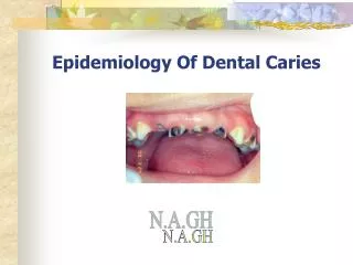
It is the most prevalent disease affecting the human race, Practically spread all over the world, Affects both sexes and all races, all socio-economic strata and people of all ages, Starts soon after teeth erupt into the oral cavity It is a paradox that teeth are the hardest tissue in the body but can be easily destroyed relatively rapidly in vivo.
What is Dental Caries? * It is a microbial disease of the calcified tissues of the teeth, characterized by demineralization of the inorganic portion and destruction of the organic substance of the tooth. * Many researches/investigations for more than a hundred years have been done, still, many aspects of the etiology of this disease is obscure and efforts at prevention are only partly successful.
A detail of a tooth (to the right = enamel). It is covered by plaque, which consists mainly of bacteria. Plaque is often found close to the gum, in between teeth, in fissures and at other "hidden" sites. Demineralization:When sugar and other fermentable carbohydrates reaches the bacteria, they form acids which start to dissolve the enamel - an early caries lesion occurs due to loss of Calcium and Phosphates Remineralization:When sugar consumption has ceased, saliva can wash away sugars and buffer the acids. Calcium and Phosphates can again enter the tooth. The process is strongly facilitated by fluorides A CAVITY occurs if the Demineralization "wins" over the Remineralization over time
The first indication of tooth decay are white spots on the enamel caused by the loss of calcium. If the demineralization process outruns the natural remineralisation process, the lesion grows and a cavity is formed. The bacteria may also produce an abscess, The bacteria may invade the pulp of the tooth, and eventually the tooth may be extracted by the dentist. causing a consistent tooth pain, especially during the night.
A tooth surface without caries. • The first signs of demineralization. • The enamel surface has broken down. • A filling has been made but the demineralization has not been stopped. • The demineralization proceeds and undermines the tooth. • The tooth has fractured.
Terminology Primary Caries: lesions on unrestored tooth surface. Secondary (recurrent) caries: lesions that developed adjacent to a filling. Residual caries: demineralized tissue that has been left behind before a filling is placed. Active caries lesion: a progressive carious lesion. Arrested (inactive) carious lesion: A lesion that may have formed years previously and then stopped further progression. White spot caries: the first sign of a caries lesion on enamel that can be detected with the naked eye. Also known as initial or incipient caries. Rampant caries: is the name given to multiple active carious lesions occurring in the same patient. This frequently involves surfaces of teeth that do not usually experience dental caries eg, bottle or nursing caries, baby caries, radiation caries, or drug-induced caries.