Download
1 / 73
740 likes | 903 Vues
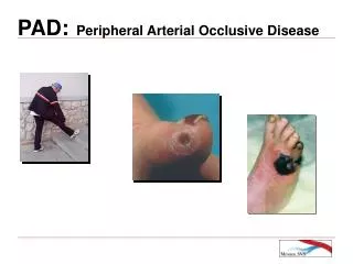
PAD vs Venous Disease: Discrepancies & Similarities Abraham Sadighi, MD, FACS GulfCoast Cardiothoracic & Vascular Surgeons Vein & Laser Treatment Center. Peripheral Arterial Disease (PAD). Disorders of the circulatory system to the extremities, viscera and head.

E N D
PAD vs Venous Disease: Discrepancies & Similarities Abraham Sadighi, MD, FACS GulfCoast Cardiothoracic & Vascular Surgeons Vein & Laser Treatment Center
Peripheral Arterial Disease (PAD) • Disorders of the circulatory system to the extremities, viscera and head
IntroductionAtherosclerotic changes Normal Artery Diseased Artery
IntroductionDisease evolution • Claudication • Rest pain • Ulceration • Gangrene • Limb loss
IntroductionMortality • Life expectancy reduced 10 years in patients with PVD • Mortality rate ~ 25% at 5 years ~ 50% at 10 years ~ 75% at 15 years • 75% of deaths caused by cardiovascular events NEJM, 1991; 325:577-8
Prevalence Total ~ 10 million U.S. patients Asymptomatic 5 million Symptomatic untreated 3.75 million Symptomatic treated 1.25 million Pentecost, et al. “Guidelines for Peripheral Percutaneous Transluminal Angioplasty of the Abdominal Aorta and Lower Extremity Vessels”; 1993
Peripheral Vascular Outcomes Limb Outcomes Population >55 yr WorseningClaudication16% Lower ExtremityBypass Surgery7% MajorAmputation4% Other CardiovascularMorbidity/Total Mortality IntermittentClaudication5% Reduce CV Risk 5-yr Mortality 30% Nonfatal Cardiovascular Event (MI/Stroke, 5-yr Rate) 20% Cardiovascular Cause 75% Adapted from Weitz JI, et al. Circulation. 1996;94:3026-3049. Prevalence Outcomes in Patients with Intermittent Claudication
Risk Factors • Tobacco abuse • Hypercholesterolemia • Hypertension • Diabetes • Obesity • Sedentary lifestyle
Risk Factors Male Gender Age (per 10 y) Diabetes Smoking Hypertension Hypercholesterolemia Fibrinogen Alcohol -4 -3 -2 -1 0 1 2 3 4 Protective Harmful Odds Ratio Belch JJF, et al. Arch Intern Med. 2003;163:884-892.
Diagnosis • Patient history • Physical examination • Laboratory values • Noninvasive vascular studies • Angiography
Patient History • Risk factors • Exercise-induced symptoms • Rest pain • Ulceration
Patient History • History—the most important aspect of the diagnostic evaluation of PAD • Location of symptoms • Description of discomfort • Exacerbating/ameliorating characteristics • Reproducible symptoms
Patient History Historical clues to the diagnosis of intermittent claudication Variable Symptom Complex Symptoms in the legs that are provoked by walking and relieved by rest Pain Aches Tiredness Tightness Soreness Weakness Numbness
Patient History Is it vascular limb pain? Historical Vascular Neurogenic Clue Etiology Etiology Onset Predictable Variable Only with walking? Yes No Relief with stopping or Yes Variable standing? Absent pedal Variable Variable pulses at rest
Patient HistoryDifferential diagnosis of PAD • Intermittent claudication • Atherosclerosis • Non-atherosclerotic • TAO/Buerger’s • PAES • CAD of the popliteal artery • FMD • Vasculitis • Neurogenic causes • Lumbar canal stenosis • Peripheral neuropathy • Venous claudication • Musculoskeletal causes • Arthritis • Bursitis • Tendonitis • Tight hamstring/quadriceps musculature • Podiatric causes • Plantar fasciitis
Physical Examination • Pulses • Bruits • Ankle-Brachial Index (ABI)
Physical ExaminationAnkle-Brachial Index • Simple, painless, accurate, highly reproducible examination • Clinically useful • Identifies patients with PAD • Major indicator of premature MI, CVA, mortality • Indications • Any patient with suspicion for PAD • Any patient at risk of PAD • Age 50 or greater with history of DM or tobacco use • Age 70 or greater regardless of risk factors
Physical ExaminationAnkle-Brachial Index • How to perform • Patient resting supine for 5-10 minutes • Continuous wave, hand-held Doppler • Measure systolic BP in both arms • Higher value is DENOMINATOR of ABI • Measure systolic BP in DP and PT • Higher value is NUMERATOR of ABI Right Arm Pressure: Left Arm Pressure: Pressure: Pressure: PT PT DP DP
Physical Examination ABI = Ankle Systolic Pressure Brachial Systolic Pressure >0.9 = Normal >0.4-0.9 = Moderate disease <0.4 = Severe disease
Physical ExaminationInterpretation and limitations of ABI ABI Interpretation Two Main Limitations Calcified ankle vessels result in artificially “normal” ABI (DM, RF) Normal ABI in patient with Aortoiliac Disease— only becomes abnormal with exercise testing Above 0.90 — Normal 0.71-0.90 — Mild Obstruction 0.41-0.70 — Moderate Obstruction 0.00-0.40 — Severe Obstruction
Physical ExaminationABI−Predictor of ischemic events CAPRIE Study ABI – inverse relationship with three-year risk of cardiovascular events and deaths 2.5 10.2% relative risk increase per 0.1 decrease in ABI(P = 0.041) 2 Risk Relative to ABPI = 1 1.5 1 0 0.2 0.4 0.6 0.8 1 ABPI Dormandy JA. Cerebrovasc Dis. 1999;9 (Suppl 1):1–128.
Noninvasive Vascular Studies • Vascular ultrasound • CT angiography • Magnetic resonance angiography
Noninvasive Vascular Studies Left SFA Stenosis CTA DSA (Pre-PTA) Right Fem-Pop BPG
Noninvasive Vascular StudiesDiagnosis−angiography Normal Abnormal
Treatment • Goals • Risk factor modification • Medical management • Minimally invasive techniques • Case studies • Surgical intervention • Follow-up care
Goals PAD Therapeutic Goals Improve functional status and quality of life Identify and treat systemic atherosclerosis Preserve the limb Prevent progression of atherosclerosis
Risk Factor Modification • Tobacco cessation • Exercise • Weight reduction • Pharmacologic intervention • Hypercholesterolemia • Hypertension • Diabetes
Medical Management Medical therapy forintermittent claudication • Symptom/Limb • Tobacco cessation • Foot care • Control of DM • Reduction in cholesterol • Antiplatelet agents • Exercise • Cilostazol • Angiogenesis • Gingko biloba • Life • Tobacco cessation • Control of DM • Reduction in cholesterol • Reduction in BP • Antiplatelet agents • Exercise
Medical ManagementPAD risk reduction therapies • Smoking • Complete cessation • Diabetes mellitus • HbA1c <7.0%, treat other risk factors • Dyslipidemia • LDL <100 mg/dL, modify HDL and TG • Hypertension • BP <140/90 or <130/80 in diabetes • ACE inhibitors • Antiplatelet therapy • Aspirin or clopidogrel
Minimally Invasive Techniques • Percutaneous transluminal angioplasty (PTA) • Stenting • Thrombolysis
Minimally Invasive Techniques Guidewire placement
Minimally Invasive Techniques Guidewire advanced past lesion
Minimally Invasive Techniques Balloon dilatation Percutaneous Transluminal Angioplasty
Minimally Invasive Techniques Stent expansion by a balloon catheter over a guidewire
Minimally Invasive Techniques Post-PTA/stent placement
Minimally Invasive Techniques Thrombolysis Post-thrombolytic infusion revealing stenosis
Case Study #1 Aorto/iliac disease
Case Study #1 Aorto/iliac disease
Case Study #1 Aorto/iliac disease
Case Study #1 Aorto/iliac disease
Case Study #1 Aorto/iliac disease
Case Study #1 Aorto/iliac disease pre-PTA stenting Aorto/iliac disease post-PTA stenting
Case Study #2 Subclavian disease
Case Study #2 Subclavian disease
Case Study #2 Subclavian disease pre-PTA stenting Subclavian disease post-PTA stenting
Case Study #3 Pre-thrombolysis Post-thrombolysis
Case Study #3 Angioplasty post-thrombolysis
Surgical Intervention • Bypass grafts • Amputation