Download
1 / 81
810 likes | 851 Vues
Learn about the anatomy of the spine, causes of low back pain, diagnostic methods, and treatment options provided by Dr. Steven Stoltz, a rheumatologist specializing in back conditions.

E N D
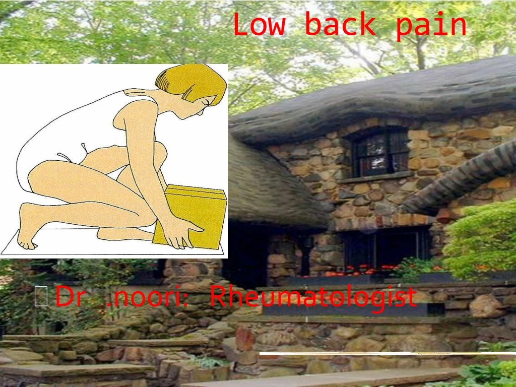
Low back pain • Dr .noori: Rheumatologist
back symptoms are the mostcommon cause of disability in those <45 years; (2) low back pain is the secondmost common reason for visiting a physician in the United States.
Anatomy The anterior portion of the spine consists of cylindrical vertebral bodies separated by intervertebral disks and held together by the anterior and posterior longitudinal ligaments. The intervertebral disks are composed of a central gelatinous nucleus pulposus surrounded by a tough cartilaginous ring, the annulus fibrosis. Disks are responsible for 25% of spinal column length and allow the bony vertebrae to move easily upon each other
nucleus pulposus and degeneration of the annulus fibrosus increase with age and results in loss of height. The disks are largest in the cervical and lumbar regions where movements of the spine are greatest. The functions of the anterior spine are to absorb the shock of body movements such as walking andrunning, and to protect the contents of the spinal canal.
The nerve roots exit at a level above their respective vertebral bodies in the cervical region (e.g., the below C7 nerve root exits at the C6-C7 level) and heir respective vertebral bodies in the thoracicand lumbar regions (e.g., the T1 nerve root exits at the T1-T2 level). The cervical nerve roots follow a short).
Causes of Low Back Pain • Lumbar “strain” or “sprain” – 70% • Degenerative changes – 10% • Herniated disk – 4% • Osteoporosis compression fractures – 4% • Spinal stenosis – 3% • Spondylolisthesis – 2% Steven Stoltz, M.D.
Causes of Low Back Pain… • Spondylolysis, diskogenic low back pain or other instability – 2% • Traumatic fracture - <1% • Congenital disease - <1% • Cancer – 0.7% • Inflammatory arthritis – 0.3% • Infections – 0.01% Steven Stoltz, M.D.
Psychiatric Disease . Many patients with CLBP have a history of psychiatric illness (depression, anxiety states), or childhoodtrauma (physical or sexual abuse) that antedates the onset of back pain. Preoperative psychological assessment has been used to exclude patients with marked psychological impairments that predict a poor surgical outcome from spine surgery.
Diagnosis by Age • 20-40 • Muscular (will also see spondylolisthesis) • spondyloarthropathy • 30-50 • Disc Herniation • >50 • OA • >60 :Spinal stenosis -/tumor/osteoprosis • Spinal stenosis
On the Job • Heavy lifting • Static posture • Bending and twisting • Vibration • Most predictive? • Psychosocial factors (monotony, job dissatisfaction, etc.)
History • Location of pain • Onset of pain • Acute, chronic. • Inflammatory or mechanical • Consistency of the pain • Constant vs. Intermittent pain • Bowel and Bladder signs • Changes in activity
Pain of spine origin may be located in the back or referred to the buttocks or legs. Diseases affecting the upper lumbar spine tend to refer pain to the lumbar region, groin, or anterior thighs. Diseases affecting the lower lumbar spine tend to produce pain referred to the buttocks, posterior thighs, or rarely the calves or feet.
The differential diagnosis :a variety of serious and treatable conditions, including epiduralabscess, fractur/hematoma,, or tumor.Fever, constant pain uninfluenced by position, sphincter abnormalities, or signs of spinal cord disease suggest an etiology other than lumbar disk disease. .
- Anatomy Lesson #1 Steven Stoltz, M.D.
STRAIGHT LEG RAISE TEST STRAIGHT LEG RAISE TEST The straight leg raise test is positive if pain in the sciatic distribution is reproduced between 30° and 70° passive flexion of the straight leg. Dorsiflexion of the foot exacerbates the pain
risk factors for serious underlying diseases; the majority of these are due to radiculopathy, fracture, tumor, infection, or referred pain from visceral structures
Red Flags • History of cancer • Unexplained weight loss • Intravenous drug use • Prolonged use of corticosteroids • Older age • Major Trauma • Osteoporosis • Fever • Back pain at rest or at night • Bowel or bladder dysfunction Steven Stoltz, M.D.
Discogenic Pain • common cause of chronic or recurrent low back and leg pain. Disk disease is most likely to occur at the L4-L5 or L5-S1 levels, but upper lumbar levels are involved occasionally.
Sciatica • The sciatic nerve is the longest nerve in your body. It runs from your spinal cord to your buttock and hip area and down the back of each leg. The term "sciatica" refers to pain that radiates along the path of this nerve — from your back down your buttock and leg.
Discogenic Pain • The cause is often unknown; the risk is increased in overweight individuals. Disk herniation is unusual prior to age 20 years and is rare in the fibrotic disks of the elderly. Genetic factors may play a role in predisposing some patients to disk disease.
The pain may be located in the low back only or referred to a leg, buttock, or hip. A sneeze, cough, may cause the nucleus pulposus to prolapse, pushing the frayed and weakened annulus posteriorly. With severe disk disease, the nucleus may protrude through the annulus (herniation) or become extruded to lie as a free fragment in the spinal canal.
. Symptoms and signs are usuallyunilateral, but bilateral involvement does occur with large central disk herniations that compress multiple roots or cause inflammation of nerve roots within the spinal canal. There is suggestive evidence that lumbar disk herniation with a nonprogressive nerve root deficit can be managed nonsurgically
Cauda equina syndrome (CES) signifies an injury of multiple lumbosacral nerve roots within the spinal canal distal to the termination of the spinal cord at L1-2. Low back pain, weakness and areflexia in the legs, saddle anesthesia, or loss of bladder function may occur.
Investigations • Note: if the diagnosis would appear to be simple back pain, then no investigationis required. • If other diagnoses are entertained, appropriate investigations are in order, depending upon the suspicion. • Imaging • X-rays- for fractures and osteoporosis • CT-scan-spondylolisthesis • Bone scan • MRI-soft tissues,disc and nerves. • Bloods • Full blood count, ESR, CRP, urine analysis if cancer, infection or inflammation suspected.9,10 • LFTsmay be helpful. Alkaline phosphatasecan be elevated in metastatic disease and Paget's disease of bone. • PSA will be raised particularly in carcinoma of the prostate. • EMG and NCV
Role of X-rays (Radiology) • Usually unnecessary and not helpful • Plain X-ray: • Age>50 years • No improvement after 6 weeks • Other worrisome findings • MRI: • After 6 weeks if have sciatica Steven Stoltz, M.D.
diagnosis of nerve root injury *****the history, examination, results of imaging studies, and the EMG are concordant. Thecorrelation between CT and EMG for localization of nerve root injury is between 65 and 73%. Up to one-third of asymptomatic adults have a lumbar disk protrusion detected by CT or MRI scans. .