Download
1 / 27
310 likes | 598 Vues
APPROACH TO VASCULAR INJURY. BY DR SIKHOSANA. Mechanisms of injury. Penetrating Blast Blunt iatrogenic. Pathophysiology . Missile damage is related to the velocity Shotgun causes multiple perforations and can cause embolization Blunt trauma results from shearing or distraction

E N D
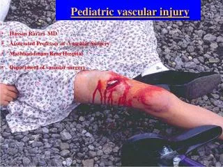
APPROACH TO VASCULAR INJURY BY DR SIKHOSANA
Mechanisms of injury • Penetrating • Blast • Blunt • iatrogenic
Pathophysiology • Missile damage is related to the velocity • Shotgun causes multiple perforations and can cause embolization • Blunt trauma results from shearing or distraction • Vascular spasm occurs at or distal to the injury due to the unapposed sympathetic constriction, it is not the cause for ischaemia
Hard signs • Pulsatile bleeding • Expanding haematoma • Thrill or bruit • Pulse deficit • ischaemia
Soft signs • History of a significant bleed • Small non expanding haematoma • Associated nerve injury • Proximity to a major vessel
Unclear presentation • Thorax injuries- suspect if there is a widened mediastinum, persistent shock, large haemothorax • Intimal injury- the pulses maybe intact but the exposed collagen is very thrombogenic
Indications for investigation: neck • Zone I and III • All gunshots • Suspicion post doppler of zone II
Mediastinum • Fracture of 1st,2nd ribs, sternum and scapula • Sterno clavicular joint dislocation • Trans axial gunshot • Widened mediastinum • Obliteration of aortic notch, left apical pleural cap, aorto-pulmonary window • Left haemothorax • Oesophageal and tracheal deviation to the right • Depression of left main bronchus
Limbs • Multiple fractures • Multiple penetrating injuries • Shotgun • Knee/elbow dislocation • Degloving injury • Gunshot tract along the long axis of the vessel
Imaging modalities • Duplex ultrasound • Angiography • CT angiography • MRA
Duplex ultrasound • Combines pulsed doppler and real time B mode ultrasound imaging • Advantages- non invasive, cheap, no radiation and sensitive • Locally used for neck zone II and single peripheral injuries
Angiography • Gold standard imaging and there is a therapeutic option, although it is invasive • Features suggestive of injury- extravasation of contrast, dilatation due to intimal injury, narrowing, occlusion, filling defects and AV fistula
CT angiography • Sensitivity and specificity of 90-100% • Advantage is that it is non invasive and rapid • Disadvantages – lack of therapeutic options, artifacts from foreign bodies, streak artifacts simulating intimal tears and the imaging of the arch not good on CT
MRA • Has good sensitivity • Not ideal due to the time taken for the investigation
Bleeding control • Pressure • balloon
Management • All vascular injuries should be repaired as ASAP to avoid delayed bleeding, compressive haematoma and limb compromise • We do not believe in conservative management of minimal arterial injuries because the history is unpredictable, poor patient compliance and too late presentation of complications
Mangled extremity severity score • Skeletal/soft tissue injury • Limb ischaemia • Shock • Age Score of >7 is accurate for predicting eventual need for amputation
Diagnostic fasciotomy • More than 6 hours presentation
Prophylatic fasciotomy • Prolonged hypotension • Extensive soft tissue injury • Arterial and venous injury • Bone plus vascular injury • Delayed vascular repair • Inability to assess the patient, e.g. head/spinal injury
Therapeutic fasciotomy • Increased tissue turgor • Extensive deep haematoma in the presence of ischaemia • FASCIOTOMY BEFORE VASCULAR REPAIR
Principles of vascular repair • Digital or sponge pressure and catheter to control bleeding • Prophylatic antibiotics • Access available to the groin for the graft • Wide exposure with proximal and distal control • Edges debrided to healthy intima • Embolectomy and flushing with heparin saline • Vascular repair before ortho • Adequate tissue cover of the vascular repair
Techniques of repair • Lateral – for wide calibre vessels • Patch- to prevent stenosis • End to end- single tethering stitch should hold and < 4mm vessel should have interrupted sutures • Interposition graft- NB similar size with the injured vessel • Ligation- gross contamination and unstable patient
Types of grafts • Vein- no cost and low infection rate • Arterial- same advantages as the vein but the donor site may need to be replaced • Synthetic- ? Higher infection risk, expensive and poor patency across joints
Causes of graft thrombosis • In flow • Anastomosis – intimal injury, adventitia, tension, stenosis, poor graft • Run off
Primary amputation • Dead leg • 2 or more dead compartments • Mangled limb
Endovascular • Embolisation • Stenting • Balloon occlusion
Conclusion • All vascular injuries should be repaired as soon as they are identified • We do not have enough man power to treat minimal injuries consevatively