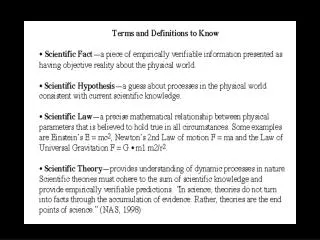
Nucleic acids
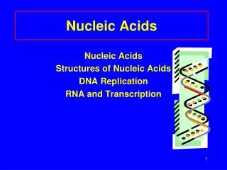
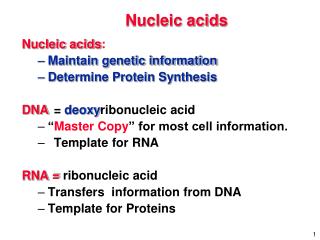
Nucleic acids. Nucleic acids : Maintain genetic information Determine Protein Synthesis DNA = deoxy ribonucleic acid “ Master Copy ” for most cell information. Template for RNA RNA = ribonucleic acid Transfers information from DNA Template for Proteins. Nucleic Acids. Chromosomes

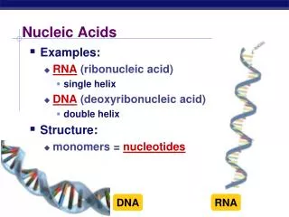
Nucleic acids
E N D
Presentation Transcript
Nucleic acids • Nucleic acids: • Maintain genetic information • Determine Protein Synthesis • DNA = deoxyribonucleic acid • “Master Copy” for most cell information. • Template for RNA • RNA = ribonucleic acid • Transfers information from DNA • Template for Proteins
Nucleic Acids • Chromosomes • (in nucleus) Have genes 1 gene 1 enzyme or protein Enzymes determine external & internal characteristics
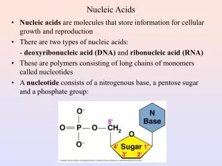
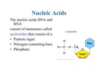
NUCLEIC ACIDS • Long chains (polymers) of repeating nucleotides. • Each nucleotide has 3 parts: A heterocyclic Amine Base A sugar A phosphate unit
Nucleotide = phosphate + sugar + base Phosphate Base Sugar -N-glycosidic linkage Nucleoside = sugar + base
Nucleic Acids • Nucleic Acids = polymers of Nucleotides. base B B B B B B P S P S P S P S P S P S phosphate sugar
O O HOCH2 HOCH2 OH OH H H H H H H H H OH OH OH H THE SUGAR PART • The major difference between RNA and DNA is the different form of sugar used. Ribose C5H10O5 in RNA DeoxyRibose C5H10O4 in DNA The difference is at carbon #2.
A double ring (6 & 5 members) A single ring (6 membered) The Nitrogenous Bases • 5 bases used fall in two classes • Purines & Pyrimidines
The Nitrogenous Bases Purines: • Pyrimidines: Adenine (A) Guanine (G) Thiamine (T) In DNA only Uracil (U) In RNA only Cytosine (C)
Primary structure Adenine (A) Similar to proteins with their peptide bonds and side groups. 5’ Guanine (G) Thymine (T) Phosphate bonds link DNA or RNA nucleotides together in a linear sequence. 3’
In 1938 William Thomas Astbury took the first fiber diffraction pictures of DNA, correctly predicting, in an article in the journal Nature, the overall dimensions of the molecule and that the nucleotide bases were stacked at intervals of 3.3Å perpendicular to its long axis. It was left, however, to Watson and Crick after the Second World War to elucidate the detailed double helical structure of DNA.
Maurice Wilkins with one of the cameras he developed specially for X-ray diffraction studies
Work on x-ray diffraction patterns by Maurice Wilkins and Rosalind Franklin in 1953, revealed that the molecule had a "helical shape“.
Rosalind Franklin is most associated with the discovery of the structure of DNA. At 26, after she had her PhD, Franklin began working in x-ray diffraction - using x-rays to create images of crystallized solids. She pioneered the use of this method in analyzing complex, unorganized matter such as large biological molecules, and not just single crystals.Franklin made marked advances in x-ray diffraction techniques with DNA. She adjusted her equipment to produce an extremely fine beam of x-rays. She extracted finer DNA fibers than ever before and arranged them in parallel bundles. And she studied the fibers' reactions to humid conditions. All of these allowed her to discover crucial keys to DNA's structure. Maurice Wilkins, her laboratory's second-in-command, shared her data, without her knowledge, with James Watson and Francis Crick, at Cambridge University, and they pulled ahead in the race, ultimately publishing the proposed structure of DNA in March, 1953.It is clear that without an unauthorized peek at Franklin's unpublished data, Watson and Crick probably would neither have published their famous paper on the structure of DNA in 1953, nor won their Nobel Prizes in 1962. Franklin did not share the Nobel Prize; she died in 1958 at the age of 37.
1953, James Watson & Francis Crick and their scale model for DNA
DNA secondary and tertiary structure • Sugar-phosphate backbone • Causes each DNA chain to coil around the outsideof the attached bases like a spiral stair case. • Base Pairing • Hydrogen bonding occurs between purines and pyrimidines. This causes two DNA strands to bond together. • adenine - thymineguanine - cytosine • Always pair together! • Results in a double helix structure.
H - N N O | | N - H N N N N O | | N - H H3C H H O | | N | N - H N N N O | | N N Base pairing and hydrogen bonding guanine cytosine thymine adenine
C G T A G C C G A T Hydrogen bonding Each base wants to form either two or three hydrogen bonds. That’s why only certain bases will form pairs.
Sugar-phosphate backbone DNA coils around outsideof attached bases like a spiral stair case. Results in a double helix structure.
Crick and Watson • (1962 Nobel Prize) • Proposed the basic structure of DNA • 2 strands wrap around each other • Strands are connected by H-bonds between the amines. • Like steps of a spiral staircase
Chromosomes • The normal number of chromosome pairs varies among the species. • Animal Pairs Plant Pairs • Man 23 Onion 8 • Cat 30 Rice 14 • Mouse 20 Rye 7 • Rabbit 22 Tomato 12 • Honeybee, White pine 12 • male 8 Adder’s 1262 • female 16 tongue fern
Role of RNA and DNA in Heredity RNA and DNA are involved in three major processes in a cell related to heredity as shown below: Replication is an important process during mitosis • Replication (DNA copies itself) • Transcription (The genetic code in DNA is rewritten into RNA and carried to the ribosomes by mRNA • Translation (tRNA carries amino acids to the ribosomes as part of protein synthesis Transcription and translation are two steps in the biosynthesis of a protein
S S P P S P P P S S P S A G T C C G T C G A DNA: Self - Replication
S S P P S P P P S S P S A G T C C G T C C G G A DNA: Self - Replication
Replication of DNA Replication occurs on both halves in opposite directions.
RNA synthesis In the first step, RNA polymerase binds to apromotor sequence on the DNA chain. This insures that transcription occurs in the correct direction. The initial reaction is to separate the two DNA strands.
RNA synthesis initiation sequence termination sequence ‘Special’ base sequences in the DNA indicate where RNA synthesis starts and stops.
RNA synthesis Once the termination sequence is reached, the new RNA molecule and the RNA synthase are released. The DNA recoils.
The messenger RNA (mRNA) move outside the nucleus to the cytoplasm where Ribosomes are anxiously awaiting their arrival. rRNA rRNA Nucleus
The messenger RNA (mRNA) move outside the nucleus to the cytoplasm where Ribosomes are anxiously awaiting their arrival. rRNA rRNA Nucleus
The messenger RNA (mRNA) move outside the nucleus to the cytoplasm where Ribosomes are anxiously awaiting their arrival. rRNA rRNA Nucleus
The messenger RNA (mRNA) move outside the nucleus to the cytoplasm where Ribosomes are anxiously awaiting their arrival. rRNA rRNA Nucleus
rRNA Ribosomal RNA – rRNA: Platform for protein synthesis. Holds mRNA in place and helps assemble proteins. rRNA
UUG AUG GCU AUG 3’ 5’ • The Ribosomes are like train stations • The mRNA is the train slowly moving through the station. rRNA Codons mRNA rRNA
Transfer RNA - tRNA = • relatively small compared to other RNA’s (70-90 bases.) • transports amino acids to site of protein synthesis.
Anticodons on t-RNA Site of amino acid attachment Point of attachment to mRNA Three base anticodon site
UUU or UUC is the codon for Phe. UUG is the codon for Leu. AUG is the codon for Met.
Codons There are two additional types of codons: Initiation AUG (same as methionine) Termination UAG, UAA, UGA A total of 64 condons are used for all amino acids and for starting and stopping. All protein synthesis starts with methionine. After the poly- peptide has been made, an enzyme removes this amino acid.
activated AA MET anticodon C A G Protein Synthesis1: Activation • Each AA is activated by reacting with an ATP • The activated AA is then attached to particular tRNA... (with the correct anticodon)
MET U C A mRNA UUG AUG GCU AUG 3’ Psite A site 5’ Translation Initiation factors ribosome unit
Ala MET C A G U C A mRNA UUG AUG GCU AUG 3’ 5’ Translation Psite A site ribosome unit
peptide bond forms Ala MET mRNA C A G U C A UUG AUG GCU AUG 3’ 5’ Translation ribosome unit
Phe peptide bond Ala A G A mRNA C A G U U C C A A G GCU U UUC UUG 3’ A 5’ Translation Met ribosome unit
peptide bond forms Ala Phe mRNA C A A G G A U C A G GCU U UUC UUG 3’ A 5’ Translation Met ribosome unit
Termination • After the last translocation (the last codon is a STOP), no more AA are added. • “Releasing factors” cleave the last AA from the tRNA • The polypeptide is complete
Recombinant DNA • Circular DNA found in bacteria • E.Coli plasmid bodies • Restriction endonucleases cleave DNA at specific genes • Result is a “sticky end” • Addition of a gene from a second organism • Spliced DNA is replaced and organism synthesizes the new protein
Recombinant DNA Bacterium Remove gene segment sticky ends DNA Plasmid Cut gene for insulin Replace in bacterium