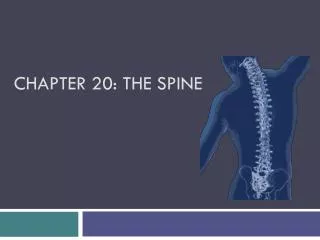
Chapter 25: The Spine
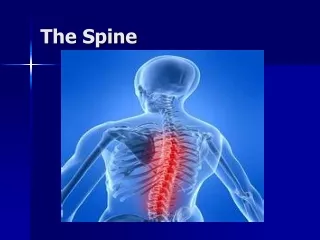
Chapter 25: The Spine. Anatomy of the Spine. Functional Anatomy of the Spine. Movements of the spine include flexion, extension, right and left lateral flexion and right and left lateral rotation Minimal movement w/in the thoracic region Movement of the spine and muscle contributions

Chapter 25: The Spine
E N D
Presentation Transcript
Functional Anatomy of the Spine • Movements of the spine include flexion, extension, right and left lateral flexion and right and left lateral rotation • Minimal movement w/in the thoracic region • Movement of the spine and muscle contributions • Superficial and deep musculature functioning and abdominal muscular functioning • Flexion and extension • Trunk rotation and lateral flexion
Prevention of Injuries to the Spine • Cervical Spine • Muscle Strengthening • Muscles of the neck resist hyperflexion, hyperextension and rotational forces • Prior to impact the athlete should brace by “bulling” the neck (isometric contraction of neck and shoulder muscles) • Varied of exercises can be used to strengthen the neck • Range of Motion • Must have full ROM to prevent injury • Can be improved through stretching
Using Correct Technique • Athletes should be taught and use correct technique to reduce the likelihood of cervical spine injuries • Avoid using head as a weapon, diving into shallow water • Lumbar Spine • Avoid Stress • Avoid unnecessary stresses and strains of daily living • Avoid postures and positions that can cause injury • Correction of Biomechanical Abnormalities • ATC should establish corrective programs based on athlete’s anomalies • Basic conditioning should emphasize trunk flexibility • Spinal extensor and abdominal musculature strength should be stressed in order to maintain proper alignment
Using Correct Lifting Techniques • Weight lifters can minimize injury of the lumbar spine by using proper technique • Incorporation of appropriate breathing techniques can also help to stabilize the spine • Weight belts can also be useful in providing added stabilization • Use of spotters when lifting • Core Stabilization • Core stabilization, dynamic abdominal bracing and maintaining neutral position can be used to increase lumbopelvic-hip stability • Increased stability helps the athlete maintain the spine and pelvis in a comfortable and acceptable mechanical position (prevents microtrauma)
Assessment of the Spine • History • Mechanism of injury (rule out spinal cord injury • What happened? Did you hit someone or did someone hit you? Did you lose consciousness • Pain in your neck? Numbness, tingling, burning? • Can you move your ankles and toes? • Do you have equal strength in both hands • Positive responses to any of these questions will necessitate extreme caution when the athlete is moved
Other general questions • Where is the pain and what kind of pain are you experiencing? • What were you doing when the pain started? • Did the pain begin immediately and how long have you had it? • Positions or movements that increase/decrease pain? • Past history of back pain • Sleep position and patterns, seated positions and postures
Observations • Body type • Postural alignments and asymmetries should be observed from all views • Assess height differences between anatomical landmarks
Cervical Spine Evaluation • Assess position of head and neck • Symmetry of shoulders (levels) • Will the athlete move the head and neck freely? • Assess active, passive and resisted ROM • Thoracic Spine Evaluation • Pain in upper back and scapular region • Cervical disk or trigger points (long thoracic nerve or suprascapular nerve involvement) • Lower thoracic region pain • Facet joint involvement • W/ deep inspiration and chin tucked to chest
Lumbar Spine and Sacroiliac Joint Observations • Coordinated movement of the low back involves the pelvis, lumbar spine and sacrum • Equal levels (shoulders and hip) • Symmetrical soft tissue structures bilaterally • Observe athlete seated, standing, supine, side-lying, and prone (leg position - contractures)
Palpation • Spinous processes • Spaces between processes - ligamentous or disk related tissue • Transverse processes • Sacrum and sacroiliac joint • Abdominal musculature and spinal musculature • Assessing for referred pain • Have athlete perform partial sit-up to determine tone and symmetry • Assess hip musculature and bony landmarks as well
Special Tests - Cervical Spine • Brachial Plexus Test • Application of pressure to head, neck and shoulders to re-create MOI • Lateral flexion of the neck w/ same side pain indicates a compression injury • Lateral flexion of the neck w/ opposite side pain indicates stretch or traction injury • Cervical Compression and Spurling’s Test • Compression of cervical spine compresses facets and spinal roots • Level of pain determines specific nerve root impingement • Spurling’s adds a rotational component to the cervical compression
Vertebral Artery Test • Athlete is supine • ATC extends,laterally bends, and rotates the c-spine in the same direction • Dizziness or nystagmus indicates occlusion of the vertebral artery • Refer to a physician for testing
Shoulder Abduction Test • Athlete places hand on top of head • A decrease in symptoms may indicate the presence of nerve root compression, due possibly to a herniated disk
Test Done in Standing Position • Forward bending • Observe movement of PSIS, test posterior spinal ligaments • Backward bending • Anterior ligaments of the spine • Disk problem • Side bending • Lumbar lesion or sacroiliac dysfunction • Standing Trunk Rotation • Assessment of symmetrical motions w/out pelvic movement
Test Done in Sitting Position • Forward bending - PSIS motions and restrictions • Rotation - lumbar spine motion symmetry • Hip Rotation - IR and ER to assess integrity and status of the piriformis muscle • “Sign of the Butt” - used to assess potentially serious hip pathology • Pain w/ passive ROM, straight leg raise, and hip flexion w/ knee flexion • Capsular pattern= limitation of flexion, abduction, internal rotation w/ slight limitations in hip extension and no limitation of external rotation • Non-capsular pattern of limitation - gross limitation in all ranges • External rotation limitation is the key motion lost in this test
Slump Test • Monitor changes in pain as sequential changes in posture occur • 1. Cervical spine flexion • 2. Knee extension • 3. Ankle dorsiflexion • 4. Neck flexion released • 5. Both legs extended • Assessment of neural tension
Tests Done in Supine Position • Straight Leg Raise • 0-30 degrees = hip problem or nerve inflammation • 30-60 degrees= sciatic nerve involvement • W/ ankle dorsiflexion = nerve root • 70-90 degrees = sacroiliac joint pathology • Kernig’s test • Unilateral straight leg raise (lumbar pain into buttocks) • Impingement of nerve root due to disk, bony entrapment or irritation of meninges • Brudzinkski’s test • Modified Kernig’s w/ neck flexion • Lumbar disk or nerve root irritation
Well Straight Leg Raising Test • Performed on the unaffected side, may produce pain in the low back on the affected side and cause radiating pain in the sciatic nerve
Milgram and Hoover Straight Leg Raising Test • Milgram test involves a bilateral straight leg raise that increases intrathecal pressure placing pressure on the disk and nerve roots • The Hoover test is a variation that utilizes a unilateral straight leg raise
Bowstring test • Used to determine sciatic nerve involvement • Leg (on affected side) is lifted until pain is felt • Knee is flexed to relieve pressure and popliteal fossa is palpated to elicit pain (along sciatic nerve) • To verify problem w/ nerve root, leg is lowered, ankle is dorsiflexed and neck is flexed. • Return of pain verifies nerve root pathology • FABER and FADIR tests • FABER or Patrick’s test is used to assess hip or SI joint dysfunction • FADIR is used to assess problems of the lumbar spine
Knee to Chest • Bilateral - increases symptoms to lumbar spine • Single - pain in posterolateral thigh may indicate problem with sacrotuberous ligament • Pulling knee to opposite shoulder that produces pain in the PSIS region may indicate sacroiliac ligament irritation • SI Compression and Distraction Tests • Used for pathologies involving SI joint
Pelvic Tilt Test • Anterior and posterior tilts that increase the pain on the side being stressed indicate irritation of the SI joint • Can also be performed from side-lying
Tests Done in Prone Position • Press-ups • While prone, push up trunk while hips remain fixed to extend the spine • Herniated disk would be apparent with radiating pain • Localized pain = conservative treatment • Generalized pain = surgery may be necessary
Reverse Straight Leg Raise • If pain occurs in low back, an L4 nerve root irritation may be present • Spring Test • Downward pressure is applied through the spinous processes of each vertebrae to assess anterior/posterior motion • Can also be performed on transverse processes to assess rotational movement • Useful to determine hypomobility or hypermobility of specific vertebral segments
Prone Knee Flexion Test • Comparison of apparent leg lengths w/ athlete prone long-lying and w/ knees flexed to 90 degrees • If there is a short side it is indicative of a posteriorly rotated SI joint • If upon flexing the knees the lengths equalize, the posteriorly rotated SI joint is indicated
Tests Done in Side-lying • Femoral Nerve Traction Test • Hip is extended and knee is flexed to 90 degrees • As the hip is extended pain occurs in the anterior thigh = nerve root impingement in the lumbar area • Posterior Rotational Stress Test • Pain on movement near PSIS indicates irritation of the SI joint • Localizes pain to a specific point - does not indicate direction of dysfunction • Piriformis Muscle Stretch Test • Flexing both hips to 90 degrees and lifting the top leg places the piriformis in a stretched position • Increasing pain indicates myofascial pain in that muscle
Iliotibial Band Stretch Test • Test will often provoke pain in the contralateral PSIS area indicating and SI problem • SI dysfunction can lead to a shortening of the IT-Band and a perpetuation or reoccurrence of the problem
Quadratus Lumborum Stretch • Use of the pillow opens the upper quadratus to palpation • Dropping the leg off the table will provide some stretch to the muscle and possibly provoking pain or demonstrating tightness
Neurological Exam • Sensation Testing • If there is nerve root compression, sensation can be disrupted
Reflex Testing • Three reflexes in the upper extremity include the biceps, brachioradialis and triceps reflexes • Tests C5, C6, and C7 nerve roots respectively • The two reflexes to be tested in the lower extremity are the patellar tendon and Achilles tendon reflexes • Used to assess the L4 and S1 nerve root respectively
Recognition and Management of Specific Injuries and Conditions
Cervical Spine Conditions • Mechanisms of Injury
Cervical Fractures • Etiology • Generally an axial load w/ some degree of cervical flexion • Signs and Symptoms • Neck point tenderness, restricted motion, cervical muscle spasm, cervical pain, pain in the chest and extremities, numbness in the trunk and or limbs, weakness in the trunk and/or limbs, loss of bladder and bowel control • Management • Treat like an unconscious athlete until otherwise rule out - use extreme care
Cervical Dislocation • Etiology • Usually the result of violent flexion and rotation of the head • Signs and Symptoms • Considerable pain, numbness, weakness, or paralysis • Unilateral dislocation causes the head to be tilted toward the dislocated side with extreme muscle tightness on the elongated side • Management • Extreme care must be used - more likely to cause spinal cord injury than a fracture
Acute Strains of the Neck and Upper Back • Etiology • Sudden turn of the head, forced flexion, extension or rotation • Generally involves upper traps, scalenes, splenius capitis and cervicis • Signs and Symptoms • Localized pain and point tenderness, restricted motion, reluctance to move the neck in any direction • Management • RICE and application of a cervical collar • Follow-up care will involve ROM exercises, isometrics which progress to a full isotonic strengthening program, cryotherapy and superficial thermotherapy, analgesic medications
Cervical Sprain (Whiplash) • Etiology • Generally the same mechanism as a strain, just move violent • Involves a snapping of the head and neck - compromising the anterior or posterior longitudinal ligament, the interspinous ligament and the supraspinous ligament • Signs and Symptoms • Similar signs and symptoms to a strain - however, they last longer • Tenderness over the transverse and spinous processes • Pain will usually arise the day after the trauma (result of muscle spasm) • Management • Rule out fracture, dislocation, disk injury or cord injury RICE for first 48-72 hours, possibly bed rest if severe enough, analgesics and NSAID’s, mechanical traction
Acute Torticollis (Wryneck) • Etiology • Pain on one side of the neck upon wakening • Result of synovial capsule impingement w/in a facet • Signs and Symptoms • Palpable point tenderness and muscle spasm, restricted ROM, muscle guarding, • Management • Variety of techniques including traction, superficial heat and cold treatments, NSAID’s • Use of a soft collar can be helpful as well
Cervical Cord and Nerve Root Injuries • Etiology • Mechanisms include, lacerations, hemorrhage (hematomyelia), contusion and shock • Can occur separately or together • Signs and Symptoms • Various degrees of paralysis impacting motor and sensory function; the level of injury determines the extent of functional deficits • Cord lesions at or above C3 result in death, while injury below C4 will allow for some return of nerve root function • Incomplete lesions can result in a number of different syndromes and conditions • Management • Handle w/ extreme caution to minimize further spinal cord damage