
BURN
E N D
Presentation Transcript
Objectives • Describe epidemiology of burn injury • Discuss causes of burn • Classify burn injury • Discuss Pathophysiology of burn • Assessment of burn patient • Describe treatment plans for burn patient by using ATLS principles • Discuss complications of burn BURN
Introduction Burn Tissue injury • thermal ( heat, cold) • electrical • Radiation • chemical • coagulative necrosis BURN
Epidemiology • 1% of the world population each year • USA ~ 2.4 million burn injuries/ yr & 10,000 death/yr • UK ~ 250,000 patients treated with burns & 700 deaths/yr. • In Kenya 5,000 deaths/yr • TZ(MNH) 10% of admission in pediatric surgical ward • ??BMC BURN
epid…… Age Scald - < 5 year of age flame, electrical & chemical burn - adult Sex domestic burn - females occupational - males Race No race predilection exists in burn injuries BURN
Risks factors Diseases e.g. epilepsy, diabetes Children< 5years; Elderly > 75 years Cold weather Occupational – electricians/industrial Alcoholism ??Low socioeconomic status BURN
High morbidity and mortality emotional & psychological BURN
Anatomy Skin The epidermis • derived from ectoderm • it can regenerate. The dermis • from mesoderm • cannot re-generate, BURN
AETIOLOGY Thermal injuries Scald Flame Flash Contact Chemical injuries Electrical injuries Radiation injuries Cold injuries BURN
classification • type /cause • body site • degree • size/extent • severity BURN
Class.. - type • Thermal burn • Scald • Flame burn • Contact burn • Flash • Electrical burn • Chemical burn • Radiation burn • Cold burn BURN
Class.. site • Facial burn • Head & neck • Trunk • Limbs • Perineal burn depth • Superficial burn • Epidemal • Dermal • Deep burn • Dermal • Full thickness • Mixed burn BURN
Class.. degree of tissue injury • First degree burn • Second degree burn • 2nd Degree Superficial (superficial Dermal) • 2nd Degree Deep (deep Dermal) • Third degree burn • Fourth degree burn BURN
Class.. Size/Extent Total body surface area (TBSA) burned severity of burn • Minor burn • Moderate burn • Major burn BURN
PATHOPHYSIOLOGY Burn injuries result in:- local response systemic response BURN
Pathophysiology…… LOCAL RESPONSE • Inflammation • Jackson zones (1947) • coagulation /necrosis • Stasis/ischaemia • hyperemia BURN
SYSTEMIC RESPONSE:- Significant burn massive release of inflammatory mediators, both in the wound and other sites. Pathophysiology…… BURN
Follow burn injury , neutrophils ,monocytes & platelets migrate into burn wound • Capillary permeability locally & in distinct organs. • Plasma oncotic pressure • Interstitial oncotic pressure due to increased capillary permeability protein loss edema in burned & un-burned tissues BURN
Biochemical … ↓ tissue perfusion tissue hypoxia anaerobic resp Pyruvate↑ lactic acid metabolic acidosis alter cellular enzymes activity BURN
Biochemical….. ↓ATP↓ Na+Ka+-ATPase ↑↑Na+ intracellular & ↑↑K+ extracellular cellular swelling hyperkalemia ↓ ECF vol. Cell death by necrosis or apoptosis BURN
CVS • ↓Myocardial contractility TNF • ↓ CO due to loss of intravascular vol, ↑ viscocity & ↓cardiac contractility. These changes, coupled with fluid loss from the burn wounds systemic hypotension & end organ hypotension MOD MOF BURN
Respiratory Inflammatory mediators →bronchoconstriction, → ARDS Pulmonary dysfunction • Inhalation injury • Aspiration • Shock • Circumferential thoracic eschar BURN
GIT • mucosal atrophy • changes in the digestive absorption • intestinal permeability Thromboxane A2 prominent mesenteric vasoconstriction ↓gut blood flow compromise gut mucosal intergrity & ↓ immune fxn • Stress (Curling’s) ulcer ( stomach & duodenum). • Acute pseudo-obstruction of the colon (Adynamic ileus) • Acute dilatation of the stomach & colon. • Acalculous cholecystitis BURN
Renal Changes BV &↓ CO RBF GFR ATN ARF BURN
CNS Changes CNS dysfunction in up to 14% of burn patients • Delirium, disorientation Hypoxia • smoke inhalation, • pulmonary edema, • pneumonia BURN
Haematological • Haemoconcentration • Anaemia • Destruction of RBC • Continual loss of RBC for 1 wk • Mild thrombocytopenia (sequestration) early, followed by thrombocytosis (2-4x > normal) by end of the 1st week Persistant thrombocytopenia associated with poor prognosis suspect sepsis • DIC with generalized bleeding can occur shock, sepsis, hypoxia, reperfusion injury BURN
Immunological Innate immunity Skin Cellular Immune Function lymphocyte function Humoral Immune Function IgG & IgA BURN
Metabolic • Ebb phase • Flow phase Catabolic phase Anabolic [recovery phase] BURN
Ebb phase Occurs during the 1st 24 hours • hypothermia • CO & O2 consumption BURN
Catabolic Phase Occurs after 24 hours of burn injury • Mediated through release of catabolic hormones [ eg, catecholamines, glucocorticoids, glucagon ] and other chemical mediators e.g. cytokines, lipid mediators. • ↑ Cardiac output • ↑ Oxygen consumption • ↑ Heat production [hyperthermia] • ↑ BMR • Hyperglycemia • Proteolysis • Peripheral lipolysis BURN
BURNSTRESS Catecolamines CORTISOL GLUCAGON Proteolysis Peripheral Lipolysis Gluconeogenesis AMINO ACIDS GLUCOSE FREE FAT ACIDS BURN
Anabolic / recovery phase Characterized by:- This phase continues for weeks to months after injury Slow re-accumulation of protein and fat BURN
ASSESSMENT OF BURN INJURY Remember • Establish cause. • Associated injuries • During escape from fire. • Explosions throw patient a distance causing internal injuries. • Electrical muscular spasms can cause fractures. • Burns in enclosed space suggest inhalational injury. • Pre-existing disease states, medication, allergies, lung sensitivities. • Establish tetanus immunization status. BURN
Clinical assessment History Physical examination General Local Systemic BURN
history Patient characteristics age , occupation History of injury Time of burn Place of burn Nature of injury Intentional Unintentional Undetermined BURN
History…. • Type of burn • Thermal • Chemical • Electrical • Radiation • Cold • Mechanism of injury • Associated injuries • Associated inhalation injuries • Associated clothing ignition • Whether first aid measures was done at the site of accident BURN
ROS • PMHx ?? Epilepsy, DM, Psychosis • FSHx ??alcohol BURN
General Exam Body weight Shock Level of consciousness Dyspnoea In pain Restless ± gasping Anaemic Dehydration BURN
Physical examination Local examination [assessment of burn wound] • Examine the wound • Body region burned • Extent of burn • Burn depth • Severity of burn Systemic examination • Cardiovascular system • Respiratory system • PA • CNS BURN
Local exam Body region • Head / neck • Upper limbs • Trunk • Lower limbs • Genitalia / Perineal areas BURN
Extent of burn Size of a Burn Injury Total Body Surface Area (TBSA) Burned Palmar Method A quick method to evaluate scattered or localized burns Client’s palm = 1 % TBSA Rule of Nines (Wallace’s) A quick method to evaluate the extent of burns Major body surface areas divided into multiples of nine Modified version for children and infants (Rule of Sevens ) Lund-Browder Method Most Accurate; based on age (growth) Can be used for the adult, children & infants BURN
Burn depth • Superficial (1st Degree) • Partial Thickness Superficial (2nd Degree) Deep ( 2nd Degree) • Full Thickness (3rd Degree) • Deep-Full Thickness (4th degree) BURN
Superficial first degree burn Epidermis Wound Appearance: • Red to pink (light skin) • Mild edema • Dry and no blistering • Pain / hypersensitivity to touch • i.e. Classic sunburn • Desquamation occurs 2-3 days Wound Healing • Wound Healing spontaneous • Duration 3 to 5 days • No scarring / other complications BURN 46
Superficial second degree burn upper 1/3 of dermis • Wound Appearance • Red to pink • Wet and weeping wounds • Thin-walled, fluid-filled blisters • Mild to moderate edema • Extremely painful • Wound Healing • In 2 weeks (spontaneous) • Minimal scarring; minor pigment discoloration may occur BURN
BURN Deep second degree burn deep dermis layer • Wound Appearance • Mottled: Red, pink, to white surface • Moist • Moderate edema • Painful; usually less severe than superficial 2nd Degree superficial. • No blisters • Wound Healing • May heal spontaneously 2-6 weeks • If so Hypertrophic scarring / formation of contractures • Wound Management: • Treatment of choice surgical excision & skin grafting
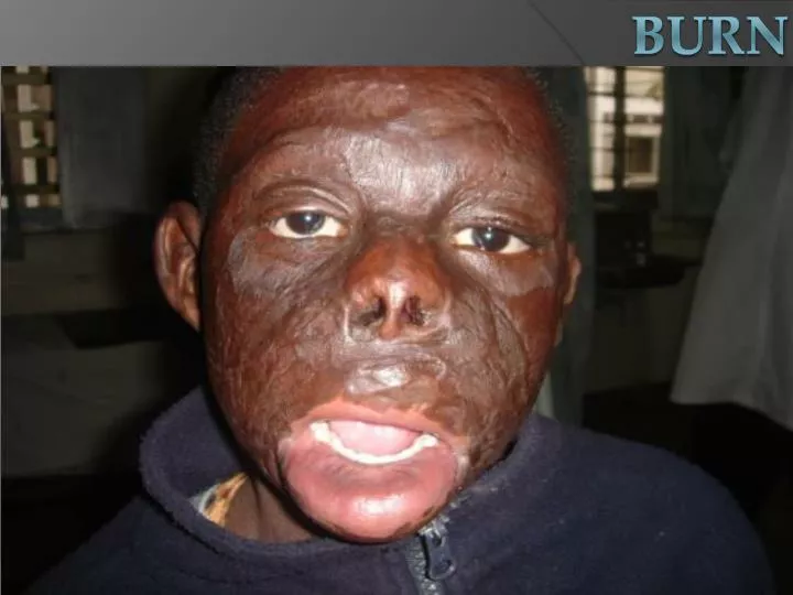
Full thickness third degree burn entire epidermis and dermisSubcutaneous fat • Wound Appearance • Dry, leathery and rigid • + Eschar (hard and in-elastic) • Red, white, yellow, brown or black • Severe edema • Painless & insensitive to palpation BURN
Wound Healing • No spontaneous healing; • No epidermal or dermal appendages remain, thus must heal by re-epithelialization from the wound edges. • Wound Management: Surgical excision & skin grafting Cx severe scarring/contracture BURN