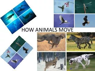
CHAPTER 30 How Animals Move
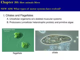
CHAPTER 30 How Animals Move. Modules 30.2 – 30.6. SKELETAL SUPPORT. 30.2 Skeletons function in support, movement, and protection. Three major functions Support Movement Protection of internal organs. Hydrostatic skeleton Exoskeleton Endoskeleton. Three main types of skeletons.

CHAPTER 30 How Animals Move
E N D
Presentation Transcript
CHAPTER 30How Animals Move Modules 30.2 – 30.6
SKELETAL SUPPORT 30.2 Skeletons function in support, movement, and protection • Three major functions • Support • Movement • Protection of internal organs
Hydrostatic skeleton • Exoskeleton • Endoskeleton • Three main types of skeletons
fluid held under pressure in a closed body compartment • cushions organs from shock • Provides body shape • Provides support for muscle action • Earthworms, hydras, and jellies have hydrostatic skeletons • Hydrostatic skeleton
The hydrostatic skeleton of a hydra Figure 30.2A
Rigid external skeleton • hard or leathery • Exoskeleton
The shells of mollusks • The exoskeleton of arthropods is made of chitin Shell (exoskeleton) Mantle Figure 30.2B, C
Most echinoderms, including sea stars and sea urchins, have an endoskeleton of hard plates beneath their skin • Endoskeleton Figure 30.2D
Vertebrate endoskeletons consist of cartilage or combo of cartilage and bone Figure 30.2E
30.5 Bones are complex living organs • Bones - several kinds of living tissues • fibrous connective tissue covers bones • Cartilage at the end of bones cushions joints • Bone tissues – surrounded by blood vessels and nerves
Cartilage Spongybone(contains redbone marrow) • A human humerus Compact bone Central cavity Yellowbone marrow Fibrousconnectivetissue Bloodvessels Cartilage Figure 30.5
MUSCLE CONTRACTION AND MOVEMENT 30.7 The skeleton and muscles interact in movement • Muscles pull on bones • Tendons: muscles to bone
Biceps contracted,triceps relaxed(extended) Tricepscontracted,biceps relaxed Biceps Biceps Triceps Triceps Tendon Figure 30.7
20.6 Muscle tissue functions in movement • Skeletal muscle - voluntary body movements • Cardiac muscle pumps blood • Smooth muscle - lines walls of internal organs ex. stomach
Unit ofmusclecontraction Nucleus Musclefiber Musclefiber Junction betweentwo cells Nucleus Muscle fiber Nucleus B. CARDIAC MUSCLE A. SKELETAL MUSCLE C. SMOOTH MUSCLE Figure 20.6
Skeletal muscle Figure 30.8
30.9 A muscle contracts when thin filaments slide across thick filaments Sarcomere Dark band Z Z Relaxedmuscle Contractingmuscle Fully contractedmuscle Figure 30.9A
Thick filament (myosin) Z line Thin filament(actin) Myosinhead ATP binds to myosin head, which is releasedfrom an actin filament. 1 Hydrolysis of ATP cocks the myosin head. 2 The myosin head attaches to an actin bindingsite. 3 The power stroke slides the actin (thin)filament toward the center of the sarcomere. 4 Figure 30.9B
30.10 Motor neurons stimulate muscle contraction • Motor neurons carry AP that initiate muscle contraction • A motor unit consists of a neuron and all the muscle fibers it controls • Strength of muscle contraction depends on number of motor units activated
Motorunit 1 Motorunit 2 Spinal cord Nerve Motor neuroncell body Motor neuronaxon Neuromuscularjunctions Nuclei Muscle fibers(cells) Muscle Tendon Bone Figure 30.10A
A neuron releases neurotransmitter acetycholine • Acetycholine triggers AP in muscle fiber • Calcium released from ER • Calcium initiates muscle contraction • neuromuscular junctions
Motor neuronaxon Action potential Mitochondrion Tubule Endoplasmicreticulum (ER) Myofibril Ca2+ releasedfrom ER Plasma membrane Sarcomere Figure 30.10B
CHAPTER 25Control of the Internal Environment Modules 25.1 – 25.4
Heat Lighttouch Pain Cold (Hair) Lighttouch Epidermis Dermis Nerve Touch Strongpressure Figure 29.3A
29.3 Specialized sensory receptors detect five categories of stimuli • Pain receptors • Sense dangerous stimuli • Thermoreceptors • Detect heat or cold • Mechanoreceptors • Respond to mechanical energy (touch, pressure, and sound)
Body Temperature • Bears don’t technically hibernate • They do enter a dormant state, when their body temperature drops by several degrees • Endotherms: • derive most of their body heat from metabolism • Ectothermic- warm themselves mainly by absorbing heat from their surroundings
Thermoregulation maintains body temperature within a tolerable range
THERMOREGULATION 25.1 Heat is gained or lost in four ways • Body temperature regulation requires adjustment to heat gained from or lost to an animal’s environment Convection Radiation Evaporation Conduction Figure 25.1
Blood flow to the skin affects heat loss Top view of shark Skin Blood vesselsof gills Artery Vein Capillary networkwithin muscle Heart Artery and veinunder the skin Dorsal aorta Figure 25.2B
Too hot /too cold • When body temp goes up: - blood vessels widen (release heat – looks flushed) - sweat glands – sweat evaporates heat • When body temp goes down: - blood vessels constrict - shivering
25.3 Behavior often affects body temperature • Basking in the sun • Sitting in the shade • Bathing • Burrowing or huddling • Migrating Figure 25.3
25.4 Reducing the metabolic rate saves energy • reduced activity and lowered metabolic rate • Hibernation in cold weather Figure 25.4