Download
1 / 14
140 likes | 168 Vues
Discover the intricacies of monosynaptic reflexes in the human body, involving sensory elements, nerve pathways, and muscle responses. Explore the components of reflex latency and variations in reflex responses.

E N D
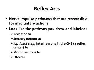
Lecture 8: Monosynaptic Reflexes • Reflex: • Common misnomer: an involuntary reaction to an external stimulus • Monosynaptic (one central synapse) • Oligosynaptic (a few central synapses; usually, 2 to 3) • Polysynaptic (many central synapses) • Tonic (slow, steady-state, maintained) • Phasic (fast, transient, in response to a change in the stimulus)
Central processing unit Efferent Afferent nerve nerve Receptor Muscle A Scheme of a Reflex A reflex arc consists of a sensory element (receptor), an afferent (sensory) nerve, a central processing unit, an efferent (command) nerve, and an effector (for example, a muscle).
Central processing unit ∆T c Efferent nerve Afferent nerve ∆T e ∆T a Muscle Muscle spindle Reaction ∆T + ∆T + ∆T a c e Time Stim Latency Reflex Latency Components of the reflex latency: • Afferent conduction delay • Central processing delay • Efferent conduction delay
a-motoneuron Efferent Ia afferents nerve Muscle Muscle spindle Monosynaptic Reflexes Monosynaptic reflexes involve one central synapse. In humans, they originate from Ia spindle afferents and induce responses in the same muscle or in muscles in the vicinity.
Central processing unit Stim Efferent Afferent nerve nerve Muscle Muscle spindle A Scheme for H-Reflex Experiments A scheme of experiments with an electrical stimulation of a muscle nerve. Note that the stimulus is applied to both afferent and efferent fibers.
EMG Time St EMG H-reflex Time St H-reflex EMG M-response Time St H-Reflex and M-Response (I) Afferent fibers are the first to react to a slowly increasing electrical stimulus. They induce a reflex muscle contraction (H-reflex). Later, efferent fibers become excited and induce a direct muscle contraction (M-response).
H-reflex EMG M-response Time St EMG M-response H-reflex Time St M-response EMG Time St H-Reflex and M-Response (II) Further increase in the strength of the stimulation leads to an increase in the M-response and suppression of the H-reflex.
AH,M M H AST Threshold Changes in the Amplitude of the H-Reflex and M-Response With the Amplitude of the Stimulus This figure shows how the peak-to-peak amplitude of the H-reflex and the M-response depends on the strength of the stimulation applied to a muscle nerve (AST). Note the nonmonotonic H-curve and a monotonic increase in the M-response.
a-motoneuron Orthodromic action potential Axon hillock Antidromic action potential Afferent fiber Efferent fiber AP Collision When an afferent fiber delivers a presynaptic action potential to an a-motoneuron whose axon hillock has just responded to an antidromic efferent action potential, the motoneuron is unable to generate another efferent action potential because of the refractory period.
1 2 3 4 5 6 EMG H-reflex M response Time St1 EMG Time St2 EMG Time St3 Effects of High Frequency Stimulation on H-Reflex Successive stimuli at a high frequency induce similar M responses but progressively smaller H-reflexes. Time scales are certainly different in the lower graphs as compared to the upper panel.
a-motoneuron Tap Muscle Spindle Tendon T-reflex EMG Time Tap Tendon Tap (T-Reflex) A tendon tap excites spindle endings and may induce a monosynaptic reflex contraction (T-reflex). Its reflex pathway is the same as for the H-reflex.
Voluntary activation a-motoneuron Ia afferents Efferent nerve Muscle spindle Muscle EMG H-reflex M response Without voluntary activation St With voluntary activation Time St H-Reflex (and T-Reflex) Under Voluntary Muscle Activation Voluntary muscle activation increases the amplitude of the H-reflex in the activated muscle through an excitation of the motoneuronal pool.
Monosynaptic Reflexes in Humans H-reflex: • Electrical stimulation of Ia afferents • Excitation of alpha-MNs through a central synapse • Efferent command to the target muscle • Twitch muscle contraction T-reflex: • Fast stretch of a muscle, leading to activation of primary muscle spindle afferents • Then same as H-reflex
a-motoneuron Antidromic action potential Orthodromic action potential Stim Muscle EMG F-wave M-response F-Wave An antidromic action potential in an efferent fiber, induced by an electrical stimulus, can induce an orthodromic action potential, leading to a muscle contraction called an F-wave.