
Tissues
Learn about the classification and functions of epithelial tissues, including their role in protection, sensory perception, secretion, absorption, and excretion. Explore epithelial tissue structures, basement membrane, and different cell shapes and layers.

Tissues
E N D
Presentation Transcript
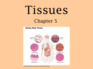
Tissues Chapter 5 http://asweknowit.net/images_edu/dwa5%20tissues.jpg
4 Types of Tissues All tissues can be classified into four major categories based on structure and function: • Epithelial: Covers and protect body surfaces, lines body cavities, moves substances in and out of blood (secretion, excretion & absorption), form glands • Connective: support, connection, transport, protection • Muscle: moves the body & its parts; specialized for contractility • Nervous: provides communication between body parts and coordinates body functions
Embryonic Development • Zygote becomes a blastocyst through mitotic division • Cells of the blastocyst regroup into primary germ layers • Endoderm, mesoderm, ectoderm • Gastrulation • Histogenesis
Epithelial Tissue Subdivided into 2 types: • Membranous • Covers the body & some of its parts • Lines body cavities (pleural, pericardial, peritoneal), blood vessels, respiratory, digestive and genitourinary tracts • Glandular • Form the secretion units of the endocrine & exocrine glands
Epithelial Tissue Functions of epithelial tissues: • Protection • Ex: skin protects body from injury & disease-causing micro-organisms • Sensory • Epithelial structures that specialize in sensory functions found in skin, nose, eye, ear • Secretion • Glandular epithelium secrete hormones, digestive juices & sweat • Absorption • Ex: gut absorbs nutrients; exchange of respiratory gases • Excretion • Ex: kidney tubules concentrate & excrete urine and other waste products
Epithelial Tissue • Basement membrane • Thin, noncellular layer of adhesive • Connects epithelial tissue and underlying connective tissue • Avascular • “without” vascular • Epithelial cells do not have blood vessels • Oxygen & nutrients diffuse from capillaries through connective tissue & basement membrane to epithelial cells
Classification of Membranous Epithelial Tissue • Cell Shape • Squamous: flat, plate-like • Cuboidal: cube-shaped; larger cytoplasm • Columnar: narrow and cylinder-shaped • Pseudostratified: single-layered; all cells touch the basement membrane but may not extend to the top of the membrane • Layers of Cells • Simple: single layer • Stratified: cells are layered on top of one another • Transitional: cell shape & layers differ
Glandular Epithelium • Specialized for secretory activity • Unicellular glands • Single celled • Ex: goblet cells • Multicellular glands • Function in clusters, solid cords or specialized follicles
Endocrine vs Exocrine • All glands are classified as endocrine or exocrine • Exocrine glands • Discharge/secrete into ducts • Ex: salivary glands • Endocrine glands • “ductless glands” • Secrete hormones directly into blood or interstitial fluid • Ex: pituitary and thyroid glands
Structural Classification of Exocrine Glands • (Table 5-2, p. 133) • Shape of gland: • Tubular • Alveolar (sac-like) • Complexity of gland: • Simple (one duct) • Compound – > 2 ducts (branched)
Functional Classification of Exocrine Glands • Apocrine • Collect secretory products at apex (tip) • Apex of cell pinches off • Cell repairs itself & repeats process • Ex: milk-producing mammary glands • Holocrine • Collect secretory product inside the cell • Rupture to release (self-destructs) • Ex: sebaceous glands (oil glands) • Merocrine • Discharge through plasma membrane • This type applies to most exocrine glands • Ex: salivary glands
Connective Tissue • Most widespread tissue in the body • Functions: • Connection • Support • Transport • Protection • Insulation
Characteristics of Connective Tissue • Common origin – mesoderm • Matrix • Intercellular material • Few cells, fibers, fluid, ground substance (material between cells) • Fibers: • Collagenous fibers • Reticular fibers • Elastic fibers
Fibers • Collagenous fibers • “white fibers” • Made of collagen (fibrous protein) • Tough, strong • Reticular fibers • Delicate • Reticulin – protein • Support small structures (ex: capillaries) • Elastic fibers • Extensible & elastic • Elastin – protein • Found in “stretchy” tissue (ex: cartilage of the external ear)
Classification of Connective Tissue • Fibrous • Loose (areolar) • Adipose • Reticular • dense • Bone • Cartilage • Hyaline • Fibrocartilage • elastic • Blood **Reference Table 5-3, pp. 134-135**
Fibrous Connective Tissue • Loose connective (areolar) tissue (fig 5-13) • Stretchable • most abundant connective tissue in the body • Connects adjacent structures • Ex: btwn other tissues and organs • Ex: superficial fascia
Fibrous Connective Tissue 2. Adipose tissue (fig 5-14) • Contains mainly fat cells • Supportive/protection pads around kidneys & other body structures • Storage deposit for excess food • Insulating material, conserves body heat
Fibrous Connective Tissue 3. Reticular Tissue (Fig 5-16) • 3D web of reticular fibers • Forms the framework of the spleen, lymph nodes & bone marrow • Meshwork filters harmful substances out of the blood
Fibrous Connective Tissue 4. Dense Fibrous Tissue (fig 5-17, 5-18, 5-19) • Densely packed fibers • Regular Dense CT • Fibers arranged in regular, parallel rows • Collagen fibers • Flexible, strong • Tendons (muscle to bone) & ligaments (bone to bone) • Irregular Dense CT • Fibers intertwine • Withstand stress from any direction • Ex: dermis (inner layer of skin); outer capsule of kidney & spleen
Bone Tissue • We will cover this when we cover the skeletal system • Just know that bone is a type of connective tissue
Cartilage • Only 1 cell type – chondrocyte • Located in lacuna • Avascular – receive nutrients via diffusion • Injuries to cartilage heal slowly due to poor nutrient delivery
Cartilage - Types • Hyaline cartilage • Most common • Covers ends of long bones (where joints articulate) • Found in supporting rings of respiratory tubes • Fibrocartilage • Strongest, most durable • Intervertebral disks • Menisci in knee joint • Elastic cartilage • Fine elastic fibers • High degree of flexibility • External ear
Blood • Unusual type of connective tissue • No ground substance • Matrix = plasma (55%) • Formed elements = blood cells (45%) • Erythrocytes – RBCs • Leukocytes – WBCs • Thrombocytes – platelets • Transport function • Respiratory gases, nutrients, waste products
Anthony’s Textbook of Anatomy and Physiology 17th Edition. Thibodeau, Gary A. PhD and Patton, Kevin T. PhD. Mosby, Inc.


