Fertilization and Sexual Differentiation
480 likes | 649 Vues
Fertilization and Sexual Differentiation. April 8 th 2010. So far have discussed ultimate causation Ontogeny (development) is an important component of proximate causation. On tap for today. Fertilization Ovulation Sperm maturation Capacitation Acrosomal Reaction Fusion of nuclei

Fertilization and Sexual Differentiation
E N D
Presentation Transcript
Fertilization and Sexual Differentiation April 8th 2010
So far have discussed ultimate causation • Ontogeny (development) is an important component of proximate causation.
On tap for today • Fertilization • Ovulation • Sperm maturation • Capacitation • Acrosomal Reaction • Fusion of nuclei • Sexual Differentiation
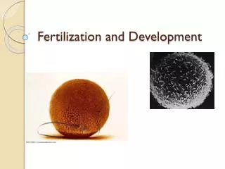
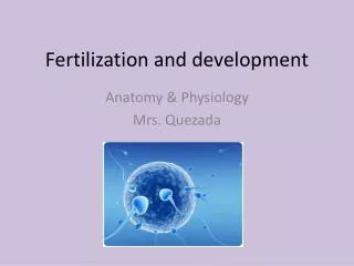
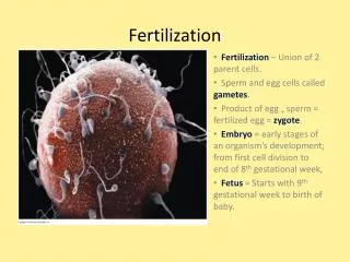
Fertilization precursors Oocyte is released along with corona radiata. Clear ring around the ovum is the zona pellucida. • Women: Ovulation Secondary oocyte released. • Still has 1 more round of meiosis • Accompanying the oocyte: • Corona radiata – follicular cells that accompanied ovum during ovulation. • Zona pellucida – adjacent to ovum. • Extracellular matrix proteins that form a barrier assuring species specific fertilization • http://www.med.uiuc.edu/histo/large/atlas/image/w63/20e1.htm Ovum with corona radiata
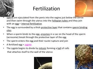
Fertilization precursors • Men: • ~10 hour journey for sperm – vagina, cervix, fallopian tube – long road. • Some sperm are held up by the folds of the cervix and are gradually released into the cervical canal; this gradual release increases the chances of fertilization. • Prostoglandins in semen stimulate muscle contractions • Only about ½ of sperm are motile. • Many don’t make it to final destination: • Fail to get through cervical mucous and don’t reach uterus • Hostile environment – uterus is acidic • Some go down wrong tube • 300 million sperm 3 million make it to uterus 2000 sperm reach the fertilization site
Sperm Morphology • Head: Acrosome and DNA • The acrosome contains enzymes which are released when sperm reaches ovum. • Acrosomal reaction enzymes digest the outer membrane of the egg, allowing penetration of the sperm. • Single set of chromosomes. • Midpiece: Mitochondria • contains numerous mitochondria. • These use sugars (fructose) in semen to generate ATP to provide the energy for tail movement. • Midpiece volume as an indicator of sexual promiscuity of females and sperm competition. • Volume is positively correlated with relative testes sizes in primates. The larger the testes (and the greater the likelihood of sperm competition), the larger the sperm midpiece. • Tail: microfilament for movement
Fertilization precursors: Capacitation • Sperm cannot fertilize oocytes when they are newly ejaculated. • Takes 5-7 hours. Various biochemical changes, especially membrane of sperm. • Sperm become “fertilization competent” • This maturation of sperm is called capacitation • Capacitation occurs in the uterus & oviducts and is facilitated by substances in the female tract. • Capacitated sperm are hyperactive. • Capacitation is required for the acrosomal reaction to take place.
Big egg, little sperm Zona Pellucida Perivitelline space
Fertilization: The acrosome reaction • The acrosome reaction: • Sperm bind to zona pellucida • (binding is species-specific) • Release of acrosome enzymes zona-digesting enzymes • Must be completed before the sperm can fuse with the secondary oocyte • The acrosome reaction provides the sperm with an enzymatic drill to get through zona pellucida • Flagella action aids penetration • Good acrosome & flagella motion are critical in male fertility
Acrosome Reaction • Proteins (red) on the sperm come into contact with the zona pellucida. • This causes the acrosome vesicle (green) to be released, spilling all of the enzymes necessary for moving towards the egg. • The actin (pink) goes from a globular state to the filamentous state pushing out the acrosomal process and exposing Bindin molecules (blue). • When the sperm cell reaches the vitelline envelope, other recognition molecules allow bindin of the correct species to attach the sperm tightly to the egg.
Acrosome reaction Sea urchin Video: http://worms.zoology.wisc.edu/urchins/SUfert_acrosome.html http://www.luc.edu/depts/biology/dev/acrosanm.htm
Fertilization • Penetration of the zona pellucida around the oocyte (takes about 20 mintues) • Once sperm penetrates zona pellucida, the zona reaction occurs: • This reaction makes the zona pellucida impermeable to other sperm (binding receptors deactivate (slow), membrane ‘hardens’(fast)) • When more than one sperm manages to enter the ovum, the fetus nearly always aborts. • Videos: • http://hthbiotech.sandiegostc.org/fertilization.html • http://arbl.cvmbs.colostate.edu/hbooks/pathphys/reprod/fert/fert.html
Fertilization • Fusion of plasma membranes of oocyte and sperm • Head and tail of a sperm enter the cytoplasm of the oocyte, but the sperm plasma membrane remains behind. • Sperm mitochondria are dispersed into the egg cytoplasm and discarded. • Egg is activated: • 2nd meiotic division of oocyte is completed. • mature ovum and polar body created (which will disintegrate and die)
Fertilization • For fertilization to occur, egg and sperm must meet between 2 – 48 hrs after sex. • Egg is only fertilizable for 48 hrs.
Cleavage • 24 hr after fertilization, the mammalian zygote (1 cell) begins cleavage. • mitotic divisions. • The egg cytoplasm is divided into smaller cells. • Progresses through 2-cell, 4-cell, 8-cell, and 16 cell stages. • The cells in cleavage stage embryos are known as blastomeres. • Soon after development of the 8-cell or 16-cell embryo (depending on the species), the blastomeres begin to form tight junctions with one another, leading to deformation of their round shape and formation of a mulberry-shaped mass of cells called a morula. 2-cell stage 16-cell stage (morula)
Growth and differentiation • Formation of junctions between blastomeres gives the embryo an outside & inside. • Get accumulation of fluid inside the embryo, which signals formation of the blastocyst. • Fluid causes it to expand. • The blastocyst stage is also a landmark in that this is the first time that two distinctive tissues are present. • Trophoblast cells – hollow sphere of cells. Will go on to contribute to fetal membrane systems. • Inner cell mass – small cluster of cells. Destined to become embryo and fetus. An early blastocyst, containing a small amount of blastocoelic fluid
Cleavage As the blastocyst expands, the zona pellucida degenerates, in preparation for implantation. The zona pellucida keeps the developing embryo from attaching to the lining of the fallopian tube until it reaches the uterus.
Blastocyst consists of: Inner cell mass (forms embryo) Blastocyst cavity Outer layer – trophoblast (forms placenta) Blastocyst attaches to uterine wall
Implantation • After zona pellucida degenerates, trophoblast cells burrow into the lining of uterus (endometrium) • The blastocyst becomes deeply embedded within the uterine lining. "Holes" in the outer trophoblast layer form and begin to fill with maternal blood. The embryo has now made its first contact with maternal blood for nourishment. • Placenta is derived from both embryonic trophoblast and maternal endometrial cells.
Gastrulation • Second week of development: gastrulation • Three important things happen: • The three primary germ layers are established. • Ectoderm gives rise to skin, sense organs, nervous system • Endoderm gives rise to digestive and respiratory tracts • Mesoderm gives rise to everything else, e.g., muscles, bone, circulatory system, etc. • The basic body plan is established, including the physical construction of the rudimentary primary body axes. • As a result of the movements of gastrulation, cells are brought into new positions, allowing them to interact with cells that were initially not near them. Allows for the development of organs and the development of the nervous system.
Embryo & Foetus • Weeks 1-3 • Fertilization, Implantation and Gastrulation • Weeks 3-8 • Embryo: • Forming major body structures, such as the head, spine, and internal organs. This is the time when most birth defects develop. • Weeks 9 – birth • The embryonic period lasts until about 8 weeks after conception (about 10 weeks from the last menstrual period). • After this point, it is called a fetus. • Lots of really neat but pretty complicated tissue differentiation. • Organ systems largely in place by 6-7 months
Sexual Determination • Chromosomal/genetic sex: XX or XY • Gonadal sex: • Males have testes and associated tubing while females have ovaries, uterus and oviducts • Hormonal sex: • Females have a high estrogen/androgen ratio, the opposite for males • Morphological sex: • Males are larger with external genitalia. Sexes have different secondary sexual characteristics at puberty • Sexual orientation: • What gender you are attracted to • Gender identity: • What gender you feel yourself to be
Chromosomes: review • Humans: 23 pairs of chromosomes • 22 pairs: autosomal • 1 pair: sex chromosomes • Y: smaller, contains genes for maleness • X: larger • Both with many genes, most not related to sex
Chromosomal sex • Determined at fertilization • XY: male • XX: female • Approximately equal numbers • Separation (disjunction) during meiosis • Two normal combinations possible • Genetic sex is determined by father • Barr bodies: condensed inactive X chromosome. Only females have them. Olympics. Magnified 10,000X
Differentiation • Differentiation: The development of structural organization whereby cell types and structures differ from one another, rather than just a growing mass of homogeneous protoplasm. • Three important points: • Multiplicity of cell types • The genetic identity of functionally dissimilar cell types • Equipotentiality and its disappearance • Sexual differentiation: the process by which the reproductive system develops.
Sexual differentiation • Differences between sexes are induced by the differential action of female and male gonads • Start undifferentiated – and stay this way until about 7 weeks after conception. • Then start to see changes typical to each sex. • Due to secretion and sensitivity to gonadal hormones: • Organizing effects: induce permanent sexual differentiation of various organs during early development • Activating effects: effects of hormones in mature organism – example: puberty
Sexual differentiation • Fertilization by an X or Y bearing sperm • No difference in development between males and females during first 6 weeks after conception. • Origin of gonads: • Germinal Ridge: • Will become somatic tissue of gonads. • Can develop into a testis or an ovary. • Primordial germ cells (PGCs): • Will become gametes • Originate outside embryo, in yolk sac • 3rd week: migrate into genital ridge, multiply
Sexual differentiation • What sex you will become is determined by the SRY gene • Sex-determining Region on Y chromosome • Leads to production of a protein called TDF (testes determining factor) • If present germinal ridge develops into a testis and you get a male • If absent an ovary forms and you get a female • Female is “default” • no genes “for” being female
Differentiation of internal systems • Every fetus, male or female, begins life with 2 separate systems that could eventually become internal reproductive organs. • Those 2 systems are: • The Wolffian system • Gives rise to the male internal anatomy: epididymis, vas deferens, seminal vesicles and prostate gland • The Mullerian system: • Gives rise to the female internal anatomy: Fallopian tubes, uterus, cervix
Differentiation into male internal genitalia • If XY, testes formation at 7 weeks • Interstitial cells of Leydig • TDF causes them to secrete testosterone (body and brain) • Maintains Wolffian system: • vas deferens: a tube conducting sperm from the testis to the urethra • seminal vesicles: lobe-type paired glands near the end of the vas deferens that contribute the gel-fraction to semen . This organ provides fructose, citric acid and proteins to nourish the sperm.
Differentiation into male internal genitalia • Degeneration of Mullerian system does not happen spontaneously. • A glycoprotein (not a steroid like testosterone) is produced in the seminiferous tubules • Mullerian inhibiting substance (MIS) • Sex cords develop • In puberty will become seminiferous tubules: contain the sperm producing cells. • Somatic cells of cords become the Sertoli cells: main function is concerned with nurturing the developing sperm
Differentiation into female internal genitalia • If XX, it has no testosterone and has no Mullerian inhibiting substance. • In the absence of testosterone, the Wolffian system disappears. • In the absence of Mullerian inhibiting substance, the Mullerian system does develop, and becomes a uterus and Fallopian tubes and vagina. • Ovary formation at 10-12 weeks • PGC’s mulitply then oogonia oocytes • Steroidogenic cells: secrete estrogen
External genitalia • Undifferentiated foetus: • Genital groove surrounded by genital fold (GF) • Forms an outgrowth: genital tubercle (GT)
Females: In absence of hormones, female structures develop: • clitoris develops from GT and labia from GF • Males: presence of Dihidroxytestosterone (DHT) causes genital groove to fuse, and the GT develops in the penis, and GF join to form the scrotum.
Atypical sexual differentiation • Chromosome abnormalities • Deficiencies in steroid production and sensitivity
Sex chromosome disorders • X0: Turner’s Syndrome (1 in 5,000) • Female, with non-functional ovaries • Administration of female hormones can induce menstruation and development of secondary sexual characteristics, which are absent otherwise. • XXY: Klinefelter’s syndrome (1 in 1,000) • Male, sparse facial and body hair, underdeveloped testes, inability to produce sperm • Breast development • Administration of testosterone produces a more masculine body and sexual characteristics.
Androgen Insensitivity Syndrome • Testes form; testosterone and anti-mullerian hormone produced (XY karyotype) • But: no internal male sex organs develop. • Body is not responding to testosterone that is produced. • Needed to maintain Wolffian system • However, Mullerian inhibiting substance (MIS) prevents development of female internal sex organs • Later, external genitalia develop as female due to absence of response to male hormones • At puberty, female body develops due to small amounts of estrogen produced by testes.
Androgen Insensitivity Syndrome An XY individual with androgen insensitivity syndrome. Despite the XY karyotype and the presence of testes, such individuals develop female secondary sex characteristics. Internally, however, these women lack the Müllerian duct derivatives and have undescended testes.
Dihydrotestoterone-deficiency (DHT) • XY, but no external male genitals at birth • DHT important in external genitalia. Internal is male tubing • Dominican Republic study. 33 s’s raised as females • At puberty, secondary male characteristics develop • Psychological development seemed to be male at this point
Congenital adrenal hyperplasia (CAH) • Increased size & # of cells in the adrenal glands • Adrenal glands produce not the normal adrenal hormone cortisol, but a substance that is structurally and functionally similar to testosterone. • Begins in perinatal period • For males, earlier onset of puberty • For females, excess androgens produced • External genitalia of male appearance
Congenital adrenal hyperplasia (CAH) • prenatal development as females • perinatal effects of androgens masculinize external anatomy • born with enlarged clitoris and fused labia - may look more male-like externally • delayed onset of menstruation due to androgens • behaviorally, “tomboys” but later most are heterosexual, some bisexual