Chapter 22: The Shoulder Complex
Chapter 22: The Shoulder Complex. Jennifer Doherty-Restrepo, MS, LAT, ATC Academic Program Director, Entry-Level ATEP Florida International University Acute Care and Injury Prevention. Introduction. The shoulder is an extremely complicated region of the body

Chapter 22: The Shoulder Complex
E N D
Presentation Transcript
Chapter 22: The Shoulder Complex Jennifer Doherty-Restrepo, MS, LAT, ATC Academic Program Director, Entry-Level ATEP Florida International University Acute Care and Injury Prevention
Introduction • The shoulder is an extremely complicated region of the body • Joint with a high degree of mobility, but, not without compromising stability • Involved in a variety of overhead activities relative to sport • Susceptible to a number of repetitive and overused type injuries
Functional Anatomy • Great mobility, limited stability • Round humeral head articulates with flat glenoid • Rotator cuff and long head of the biceps provide dynamic stability during overhead motion • Supraspinatus compresses the humeral head • Other rotator cuff muscles depress the humeral head Integration of the capsule and rotator cuff • Scapula stabilizing muscles also provide dynamic stability • Relationship with the other joints of the shoulder complex and the G-H joint is critical
Functional Anatomy • Scapulohumeral Rhythm • Movement of scapula relative to the humerus • Initial 30 degrees of G-H abduction • Does not incorporate scapular motion • Setting phase • 30 to 90 degrees of G-H abduction • Scapula abducts and upwardly rotates 1 degree for every 2 degrees of humeral elevation • Above 90 degrees of G-H abduction • Scapula and humerus move in 1:1 ratio
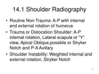
Specific Injuries • Clavicular Fractures • Etiology • MOI = fall on outstretched arm, fall on tip of shoulder, or direct impact • Occurs primarily in middle third • Signs and Symptoms • Athlete supports arm, head tilted towards injured side with chin turned away • Clavicle may appear lower • Palpation reveals pain, swelling, deformity, and point tenderness
Clavicular Fractures (continued) • Management • Closed reduction - sling and swathe immediately • Refer for X-ray • Immobilize with brace for 6-8 weeks • After removal of brace, rehabilitation includes: • Joint mobilizations • Isometric exercises • Use of a sling for 3-4 weeks • May require surgical treatment
Specific Injuries • Scapular Fractures • Etiology • MOI = direct impact or force transmitted up through humerus • Signs and Symptoms • Pain during shoulder movement • Swelling and point tenderness • Management • Sling immediately and refer for X-ray • Use sling for 3 weeks then begin PRE exercises
Specific Injuries • Fractures of the Humerus • Etiology • MOI = direct impact, force transmitted up through humerus, or fall on outstretched arm • Proximal fractures occur due to direct blow • Dislocations occur due to fall on outstretched arm • Epiphyseal fractures are more common in young athletes and occur due to direct blow or indirect blow traveling along long axis of humerus
Specific Injuries • Fractures of the Humerus (continued) • Signs and Symptoms • Pain, swelling, point tenderness, decreased ROM • Management • Immediate application of splint • Refer for X-ray • Treat for shock
Specific Injuries • Sternoclavicular Sprain • Etiology • MOI = indirect force or blunt trauma • Signs and Symptoms • Grade 1 - pain and slight disability • Grade 2 - pain, subluxation deformity, swelling, point tenderness, and decreased ROM • Grade 3 - gross deformity (dislocation), pain, swelling, and decreased ROM • Possibly life-threatening if dislocates posteriorly
Specific Injuries • Sternoclavicular Sprain (continued) • Management • RICE • Refer for reduction if necessary • Immobilize for 3-5 weeks • After immobilization period, begin PRE exercises
Specific Injuries • Acromioclavicular Sprain • Etiology • MOI = direct blow (from any direction) or upward force from the humerus • Graded from 1 - 6 according to severity of injury • Signs and Symptoms • Grade 1 - point tenderness, pain with movement • No disruption of AC joint • Grade 2 - tear or rupture of AC ligament, pain, point tenderness, and decreased ROM (abd/add) • Partial displacement of lateral end of clavicle
Acromioclavicular Sprain (continued) • Signs and Symptoms • Grade 3 - rupture of AC and CC ligaments • AC joint separation • Grade 4 - posterior dislocation of clavicle • Grade 5 – rupture of AC and CC ligaments, tearing of deltoid and trapezius attachments, gross deformity, severe pain, decreased ROM • Grade 6 - displacement of clavicle behind the coracobrachialis
Acromioclavicular Sprain (continued) • Management • Ice, sling and swathe • Referral to physician • Grades 1 – 3: non-operative treatment • 1 - 2 weeks of immobilization • Grades 4 – 6: surgery required • Aggressive rehab is required for all AC sprains • Joint mobilizations, flexibility exercises, and PRE exercises should occur immediately • Progress as tolerated – no pain and no additional swelling • Padding and protection may be required until pain-free ROM returns
A: Grade 1 • B: Grade 2 • C: Grade 3 • D: Grade 4 • E: Grade 5 • F: Grade 6
Specific Injuries • Glenohumeral Joint Sprain • Etiology • MOI = forced abduction and/or external rotation; or a direct blow • Signs and Symptoms • Pain during movement • Especially when re-creating the MOI • Decreased ROM • Point tenderness
Specific Injuries • Glenohumeral Joint Sprain (continued) • Management • RICE for 24-48 hours • Sling • After hemorrhaging subsides, modalities may be utilized along with PROM and AROM exercises to regain full ROM • When full ROM achieved without pain, PRE exercises can be initiated • Must be aware of potential development of chronic conditions (instability)
Specific Injuries • Acute Subluxations and Dislocations • Etiology • Subluxation = excessive translation of humeral head without complete separation from joint • Anterior dislocation = results from an anterior force on the shoulder with forced ABD and ER • Posterior dislocation = results from forced ADD and IR, or, falling on an extended and internally rotated shoulder
Specific Injuries • Acute Subluxations and Dislocations (continued) • Signs and Symptoms • Anterior dislocation - flattened deltoid; prominent humeral head in axilla; arm carried in slight ABD and ER rotation; moderate pain and disability • Posterior dislocation - severe pain and disability; arm carried in ADD and IR; prominent acromion and coracoid process; limited ER and elevation
Acute Subluxations and Dislocations (continued) • Management • Sling and swathe and refer for reduction • Immobilize for 3 weeks following reduction • Perform isometrics while in sling • After immobilization period, begin PRE exercises as pain allows • Protective bracing when return to play
Possible Complications of Shoulder Dislocations • Brachial nerves and vessels may be compromised • Rotator cuff injuries • Fractures • Bicipital tendon subluxation • Transverse ligament rupture
Specific Injuries • Chronic Recurrent Instabilities • Etiology • MOI = traumatic, microtraumatic (repetitive overuse), atraumatic, congenital, and neuromuscular • As supporting tissue become more lax, mobility increases • Results in damage to other soft tissue structures
Specific Injuries • Chronic Recurrent Instabilities (continued) • Signs and Symptoms • Anterior - may have clicking or pain; complain of dead arm during cocking phase (when throwing); pain posteriorly; possible impingement; positive apprehension test • Posterior - possible impingement; loss IR; crepitation; increased laxity; pain anteriorly and posteriorly • Multidirectional - inferior laxity; positive sulcus sign; pain and clicking with arm at side; possible signs and symptoms associated with anterior and posterior instability
Chronic Recurrent Instabilities (continued) • Management • Conservative treatment involves extensive strengthening of the rotator cuff and scapula stabilizers • Should be pursued before surgery is considered • Avoid joint mobilizations and ROM exercises • Various braces can be used to limit motion • Surgical stabilization may be required to improve function and comfort
Specific Injuries • Shoulder Impingement Syndrome • Etiology • Mechanical compression of supraspinatus tendon, subacromial bursa, and long head of biceps tendon due to decreased space under coracoacromial arch • MOI = overhead repetitive activities • Exacerbating factors • Laxity and inflammation • Postural mal-alignments • Kyphosis and/or rounded shoulders
Shoulder Impingement Syndrome (continued) • Signs and Symptoms • Diffuse pain • Increased pain with palpation of subacromial space • Decreased strength of external rotators compared to internal rotators • Tightness in posterior and inferior capsule • Positive impingement and empty can tests
Neer’s progressive stages of shoulder impingement… • Stage I • Result of supraspinatus or biceps tendon injury • Presents with point tenderness; pain with ABD and resisted supination with external rotation; edema; thickening of rotator cuff and bursa • Occurs in athletes < 25 years old
Neer’s progressive stages of shoulder impingement… • Stage II • Permanent thickening and fibrosis of supraspinatus and biceps tendon • Presents with aching during activity that worsens at night • May experience restricted arm motion
Neer’s progressive stages of shoulder impingement… • Stage III • History of shoulder problems and pain • Tendon defect (less than 3/8 of an inch) or possible muscle tear • Permanent scar tissue and thickening of rotator cuff • Occurs in athletes 25 - 40 years old
Neer’s progressive stages of shoulder impingement… • Stage IV • Infraspinatus and supraspinatus atrophy • Presents with pain during ABD, limited AROM and PROM, weak RROM • Tendon defect (greater than 3/8 of an inch) • Clavicle degeneration
Specific Injuries • Rotator cuff tear • Etiology • Occurs near insertion on greater tuberosity • Involve supraspinatus or rupture of other rotator cuff tendons • Partial or complete thickness tear • Full thickness tears usually occur in athletes with a long history of rotator cuff pathology • Generally does not occur in athlete under age 40 • MOI = acute trauma or impingement • Signs and Symptoms • Pain and weakness with shoulder ABD and IR • Point tenderness
Rotator cuff tear (continued) • Management • NSAID’s and analgesics • Modalities • Electrical stimulation for pain • Ultrasound for inflammation • Restore appropriate mechanics by strengthening rotator cuff to depress and compress humeral head to restore subacromial space • Severe cases may require rest, immobilization, and surgery
Specific Injuries • Shoulder Bursitis • Etiology • Chronic inflammatory condition resulting from fibrosis or fluid build-up • MOI = direct trauma or overuse • Usually occurs in the subacromial bursa • Signs and Symptoms • Pain with motion, pain during palpation of subacromial space • Positive impingement tests
Shoulder Bursitis • Management • Reduce inflammation • Cold, ultrasound, NSAID’s • Remove mechanisms precipitating condition • Maintain full ROM to reduce the risk of contractures and adhesions forming
Specific Injuries • Frozen Shoulder (Adhesive Capsulitis) • Etiology • Contracted and thickened joint capsule with little synovial fluid • Chronic inflammation resulting in contracted, inelastic rotator cuff muscles • Signs and Symptoms • Pain in all directions both with AROM and PROM • Patient resists moving the shoulder due to pain
Specific Injuries • Frozen Shoulder (continued) • Management • Aggressive joint mobilizations • Stretching of tight musculature • Electrical stimulation for pain control • Ultrasound for deep heating
Specific Injuries • Thoracic Outlet Compression • Etiology • Compression of brachial plexus, subclavian artery and vein • Due to • 1) decreased space between clavicle and first rib, • 2) scalene compression, • 3) compression by pectoralis minor, or • 4) presence of cervical rib
Thoracic Outlet Compression (continued) • Signs and Symptoms • Paresthesia, pain, sensation of cold, impaired circulation, muscle weakness, muscle atrophy, and radial nerve palsy • Positive anterior scalene test, costoclavicular test, and hyperabduction test • Management • Conservative treatment - correct anatomical condition through stretching (pec minor and scalenes) and strengthening (trapezius, rhomboids, serratus anterior, erector spinae)
Specific Injuries • Biceps Brachii Rupture • Etiology • Generally occurs near origin of muscle at bicipital groove • MOI = powerful contraction
Biceps Brachii Rupture (continued) • Signs and Symptoms • Audible snap with sudden and intense pain • Protruding bulge may appear near middle of biceps • Weakness with elbow flexion and supination • Management • Ice for hemorrhaging • Immobilize with a sling and refer to physician • Athletes will require surgery