Download
1 / 19
190 likes | 307 Vues
Effective care and removal of indwelling catheters are vital in preventing catheter-associated urinary tract infections (CAUTI). Regular perineal hygiene and monitoring urine output are crucial steps. Prior to removal, ensure the catheter balloon is fully deflated to minimize urethral trauma. After removal, monitor voiding for 24-48 hours, recording frequency and volume to ensure adequate bladder emptying. Patient education on catheter hygiene and UTI symptoms is essential for optimal recovery and self-care.

E N D
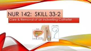
NUR 142: SKILL 33-2 Care & Removal of an Indwelling Catheter
SKILL 33-2: CARE & REMOVAL OF AN INDWELLING CATHETER • Providing regular perineal hygiene, preventing catheter-related trauma, and removing indwelling catheters as soon as possible are important interventions to reduce risk of catheter-associated urinary tract infection. • Prolonged indwelling catheterization is a major risk factor for CAUTI – Catheter Associated Urinary Tract Infection. • When removing an indwelling catheter, it is important to ensure that the catheter balloon is fully deflated to minimize trauma to the urethra.
CONTINUATION of introduction • All patient should have their voiding monitored after catheter removal for at least 24 to 48 hours by using a voiding record or bladder diary. • Record the time and amount of each voiding, including any incontinence, in the diary. • Use a bladder scan to monitor bladder function by measuring postvoid residual. • Abdominal pain and distention, a sensation of incomplete emptying, incontinence, constant dribbling of urine, and voiding in very small amounts can indicate inadequate bladder emptying requiring intervention. • The risk of urinary tract infection (UTI) increases with the use of an indwelling catheter.
ASSESSMENT • 1. Catheter Care: • A. Observe urinary output and urine characteristics. • Sudden decrease in urine output may indicate occlusion of the catheter. Cloudy, foul-smelling urine associated with other systemic symptoms may indicate CAUTI. • B. Assess for history or presence of bowel incontinence. • C. Observe for any discharge, redness, bleeding, or presence of tissue trauma round the urethral meatus (this may be deferred until catheter care). • D. Assess patient’s knowledge of catheter care.
ASSESSMENT: CONT’D • 2. Catheter Removal: • A. Review patient’s medical record, including health care provider’s order and nurses’ notes. Note length of time catheter was in place. • B. Assess patient’s knowledge and prior experience with catheter removal. • C. Assess urine color, clarity, odor and amount. Note any urethral discharge, irritation of genital region, or trauma to urinary meatus (this may be deferred until just before the removal. • D. Determine the size of the catheter inflation balloon by looking at the balloon inflation valve
PLANNING • Expected outcomes following catheter care: • Genital area is free of secretions, fecal matter, and irritation. • Patient verbalizes feelings of comfort. • Expected outcomes after catheter removal: • Patient voids at least 150 mL with each voiding no more than 6 to 8 hours after removal. • Patient verbalizes feelings of complete bladder emptying and absence of discomfort. • Patient identifies signs & symptoms of UTI – This indicates patient learning. • Explain procedure to the patient. Discuss signs and symptoms of UTI. If applicable, teach the patient how to perform catheter hygiene.
IMPLEMENTATION • 1. Identify patient using two identifiers • 2. Close room door and bedside curtain • 3. Perform hand hygiene • 4. Raise bed to appropriate working height. If side rails are raised, lower side rail on working side. • 5. Organize equipment for perineal care and/or removal of catheter. • 6. Position patient with waterproof pad under buttocks and cover with bath blanket, exposing only genital area and catheter. • A. Female in dorsal recumbent position. • B. Male n supine position.
IMPLEMENTATION – CONT’D • 7. Apply clean gloves • 8. Remove catheter securement device while maintaining connection with drainage tubing. • 9. Catheter care: • A. Female: Use non-dominant hand to gently separate labia to fully expose urethral meatus and catheter. Maintain position of hand throughout procedure. • B. Male: Use non-dominant hand to retract foreskin if not circumcised and hold penis at shaft just below glans. Maintain hand position throughout procedure. • C. Grasp catheter with two fingers to stabilize it. • D. Assess urethral meatus and surrounding tissue for inflammation, swelling, discharge, or tissue trauma and ask patient if burning or discomfort is present.
IMPLEMENTATION – CONT’D • E. Provide perineal hygiene using mild soap & warm water (use chapter 17). • F. Using clean washcloth, clean catheter. • 1. Starting close to urinary meatus, clean catheter in circular motion along its length for about 10 cm (4 inches), moving away from body. Remove all traces of soap. For male patients: Reduce or reposition foreskin after care. • G. Reapply catheter securement device. Allow slack in catheter so movement does not create tension on it.
IMPLEMENTATION – CONT’D • 10. Routinely check drainage tubing and bag. • A. Catheter is secured to upper thigh (for women) or abdomen (for men). • B. Tubing is coiled and secured onto bed linen. • C. Tubing is not looped or positioned above level of the bladder. • D. Tubing is not kinked or clamped. • E. Drainage bag is positioned below level of bladder wit hurine flowing freely into the bag. • F. Drainage bag is not overfull. Empty drainage bag when ½ full.
IMPLEMENTATION – CONT’D • 11. Catheter Removal: • (Follow Steps 1 to 10 before catheter removal.) • A. Move syringe plunger up and down to loosen and then pull back plunger to 0.5 mL. Insert hub of syringe into inflation valve (balloon port). Allow balloon fluid to drain into syringe by gravity. Syringe should fill. Make sure that entire amount of fluid is removed by comparing removed amount to volume needed for inflation. • B. Pull catheter out smoothly and slowly. Examine it to ensure that it is whole. Catheter should slide out easily. Do not use force. If you note any resistance, repeat Step 11A to remove remaining water. • C. Wrap contaminated catheter in waterproof pad. Unhook collection bag and drainage tubing from the bed.
IMPLEMENTATION – CONT’D • D. Reposition patient as necessary. Provide hygiene as needed. Lower level of bed and position side rails accordingly. • E. Empty, measure, and record urine present in drainage bag (see chapter 6). • F. Encourage patient to maintain or increase fluid intake (unless contraindicated). • G. Initiate voiding record or bladder diary. Instruct patient to tell you when need to empty bladder occurs and that all urine needs to be measured. Make sure that the patient understands how to use a collection container. • H. Explain that many patients experience mild burning, discomfort, or small-volume voiding with first voiding, which soon subsides.
IMPLEMENTATION – CONT’D • I. Inform patient to report any signs of UTI. • J. Place urine “hat” on toilet set if patient is using the toilet. Place call bell within easy reach. • 12. Dispose of all contaminated supplies in appropriate receptacle, remove gloves, and perform hand hygiene. • EVALUATION: • 1. Inspect catheter and genital area for soiling, irritation, and skin breakdown. Ask patient about any type of discomfort. • 2. Observe time and measure amount of first voiding after catheter removal – this indicates return of bladder function after the catheter is removed. • 3. Evaluate the patient for signs and symptoms of UTI. – Any patient who has recently had a catheter removed is at risk for a UTI.
UNEXPECTED OUTCOMES: • 1. Water from inflation balloon does not return into the syringe. • Reposition the patient; ensure that the catheter is not pinched or kinked. • Remove syringe. Attach new syringe and allow enough time for passive emptying. • Attempt to empty balloon by gently pulling back on the syringe plunger. • If catheter balloon does not deflate, do not cut balloon inflation valve to drain water. Notify health care provider. • 2. Patient has a fever, chills, burning, flank pain, back pain, hematuria, painful urination, urgency, frequency, lower abdominal pain, change in mental status and lethargy. • Assess for bladder distention and tenderness. • Monitor vital signs and urine output • Report findings to health care provider; signs and symptoms may indicate UTI
UNEXPECTED OUTCOMES – CONT’D • 3. Patient is unable to void after catheter removal, has sensation of not emptying, strains to void, or experiences small voiding amounts with increasing frequency. • Assess for bladder distention • Help to normal position for voiding and provide privacy • Perform bladder ultrasound (see procedural guideline 33-2) to assess for excessive urine volume in the bladder • If patient is unable to void within 6 to 8 hours of the catheter removal and/or experiences abdominal pain, notify the health care provider.
RECORDING & REPORTING • Record time for catheter care and appearance of urine; describe condition of meatus and catheter. • Record and report time of catheter removal;, amount of water removed from balloon, condition of urethral meatus and catheter, and the time, amount, and characteristics of first voided urine. • Record teaching related to catheter care, catheter removal, and fluid intake. • Report hematuria, dysuria, inability or difficulty voiding and any new incontinence after a catheter is removed.
SPECIAL CONSIDERATIONS - TEACHING • Unless contraindicated, patients with a catheter should drink at least 2200 mL of fluid per day to promote continuous flushing of the bladder and prevent sediment from collecting in the catheter tubing. • Instruct the patient to hold the collection bag below the level of the bladder when ambulating. • Instruct the patient not to disconnect the catheter from the collection tubing and the bag.
END OF SKILL: 33-2: Care & removal of an indwelling catheter • This is the end of the skill • Your book has provided a video for this skill, which is as follows: • http://booksite.Elsevier.com/Perry-Potter/ClinicalSkills/video40.php • Elsevier: Perry-Potter: Clinical Nursing Skills and Techniques, 8e – 33.2 Care of an Indwelling Catheter.