Download

1 / 20
200 likes | 224 Vues
Explore the mechanisms of polarity in cells using mathematical modeling of protein localization, focusing on the Min protein system in E. coli cell division and planar cell polarity in multicellular organisms like Drosophila. Learn the role of FtsZ, MinD, MinE, and other proteins in determining cell orientation within tissues and how interactions between proteins lead to asymmetric distributions. Discover the implications for understanding phenomena like cellular mutations and pattern formation, as explained in scientific studies.

E N D
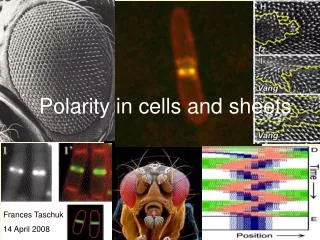
Polarity in cells and sheets Frances Taschuk 14 April 2008
E. coli cell division • Like many other prokaryotes, E. coli cells reproduce by binary fission • The plane of division is determined by the location of a ring of FtsZ protein • So how does FtsZ end up in the middle of the cell?
Modeling Min protein locations • Localization of FtsZ determined by Min protein system – MinC inhibits FtsZ polymerization • Min protein localization involves polar oscillations – modeled by Meinhardt and de Boer • Nucleoid occlusion also contributes to localization
The Min proteins • MinD – ATPase on cytoplasmic side of membrane • Recruits MinC and MinE from cytoplasm to membrane • MinE – displaces MinD from membrane – binds at flank of MinD accumulation • (MinC – inhibits FtsZ polymerization)
Oscillation of MinC/D On average, MinC concentration is highest at each end of cell
Modeling oscillations • Reaction-diffusion model using local self-enhancement and long range antagonism • Assumptions: • FtsZ, MinD, MinE produced at constant rate • All 3 diffuse rapidly • All associate with membrane by self-enhancing process • MinE displaces MinD • (not stated specifically in paper) Colocalization of MinC with MinD – ie, MinD treated as inhibiting FtsZ
Simulation and Results Calculate numerical solutions by turning these into difference equations, eg: FtsZ – blue MinD – green MinE - pink
Start from homogeneous state • http://www.pnas.org/cgi/content/full/98/25/14202/DC1/8
Consistent with observations of extended FtsZ- filaments MinD-GFP localization
What about sporulation? • Bacillus subtilis produces endospores through an asymmetrical division • Additional influence of SpoIIE protein causes FtsZ to spiral to separate rings near cell poles • One is chosen for division – mechanism unknown
Multicellular systems • Cells in multicellular organisms must organize their individual polarity to form higher-order structures • Cell polarity: apical vs basal-lateral orientation • Planar cell polarity: cell orientation within a sheet such as the epithelium
Drosophila as model system • Displays planar cell polarity in back bristles, wing hairs, and photoreceptors of the eye
Mathematical modeling of wing cell polarity • In Science, 2005 • Signaling between cells is contact-dependent • The authors propose that enough is known about the proteins involved to explain phenomena such as domineering nonautonomy. • Can be modeled as a reaction-diffusion system using partial differential equations
The feedback loop • Loop amplifies initial asymmetry, resulting in polarized distributions of planar cell polarity proteins • Fz recruits Dsh to membrane, Pk and Vang to adjacent cell’s membrane. • In each cell, Pk and Vang block local recruitment of Fz/Dsh Fz = frizzled Dsh = dishevelled Pk = Prickle-spiny-legs Vang = Van Gogh/strabismus
System of 10 nonlinear partial differential equations representing proteins and complexes Parameters unknown, so chose ones that produced certain hair pattern phenotypes - not highly sensitive to precise values Includes directional bias – actual mechanism unknown The model
Showed localization to correct membrane Able to explain autonomous mutations vs nonautonomous domineering mutations Autonomous: cells with abnormal Dsh or some abnormal Fz functions do not affect polarity of nearby cells Nonautonomous domineering: mutant Fz unable to recruit Vang to adjacent cell Results
Autonomy of mutations fzR52 – nonautonomous – does not recruit Vang-YFP fzF31 – autonomous – Fz still recruits Vang-YFP
References Meinhardt,H., de Boer, P. A. J. 2001. Pattern formation in Escehericihia coli: a model for the pole-to-pole oscillations of Min proteins and the localization of the division site. PNAS 98:25 14202-14207. Amonlirdviman, K, et al. 2005. Mathematical modeling of planar cell polarity to understand domineering nonautonomy. Science 307, 423-424. Images: http://www.nature.com/nrm/journal/v6/n11/images/nrm1745-f1.jpg http://www.nature.com/nrm/journal/v6/n11/images/nrm1745-f3.jpg http://www.nature.com/nrmicro/journal/v3/n12/images/nrmicro1290-f1.jpg http://www.pnas.org/cgi/reprint/96/9/4971.pdf http://www.pnas.org/cgi/content/full/98/25/14202/DC1/6 http://www.nature.com/nrmicro/journal/v1/n2/images/nrmicro750-f1.gif http://dev.biologists.org/cgi/content/full/129/11/2749/FIG2 http://www.mshri.on.ca/mcneill/planar.html http://web.wi.mit.edu/rebay/pub/research/images/wteye.jpg http://www.bohemianscientist.org/images/blog07/03/drosophila.jpg