Download
1 / 63
640 likes | 833 Vues
Suggested Readings: Transcription Voet and Voet: Ch 31 Watson: Ch12 pp. 437-362. Central Dogma:. RTase .

E N D
Suggested Readings: Transcription Voet and Voet: Ch 31 Watson: Ch12 pp. 437-362
Central Dogma: RTase
Figure 31-14 The two possible modes of RNA chain growth. Growth may occur (a) by the addition of nucleotides to the 3¢ end and (b) by the addition of nucleotides to the 5¢ end. Page 1226
Rifampicin inhibits prokaryotic RNA POL by binding to the subunit of RNA POL
Figure 31-17 X-Ray structure of actinomycin D in complex with a duplex DNA of self-complementary sequence d(GAAGCTTC).Intercalates and inhibits both DNA and RNA Pol. Page 1229
Figure 30-10 Schematic diagram for the nucleotidyl transferase mechanism of DNA polymerases. Page 1143
Mechanism for DNA pol Similar for RNA pol
RNA Pol E. coli DNA Pol I Klenow Fragment E. coli
Figure 31-11aX-Ray structure of Taq RNAP core enzyme. a subunits are yellow and green, b subunit is cyan, b¢ subunit is pink, w subunit is gray. Page 1224
Figure 31-12a The sequence of a fork-junction promoter DNA fragment. Numbers are relative to the transcription start site, +1. Page 1225
Figure 31-12b X-Ray structure of Taq holoenzyme in complex with a fork-junction promoter DNA fragment. Page 1225
Figure 31-10 The sense (nontemplate) strand sequences of selected E. coli promoters. Page 1223
Figure 31-13bModel of the open (Rpo) complex of Taq RNAP with promoter-containing DNA showing the transcription bubble and the active site. Page 1225
Figure 31-13aModel of the closed (RPc) complex of Taq RNAP with promoter-containing DNA extending between positions –60 and +25. Page 1225
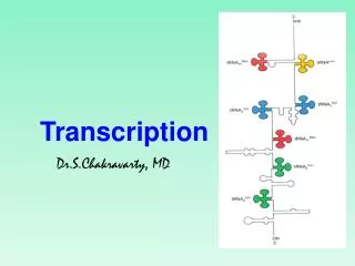
Figure 31-15 RNA chain elongation by RNA polymerase. Page 1227
Figure 31-16 An electron micrograph of three contiguous ribosomal genes from oocytes of the salamander Pleurodeles waltl undergoing transcription. Page 1228