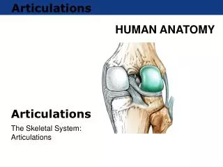
Articulations
Articulations. The place of union or junction between two or more bones of the skeleton (also between cartilage and bones/teeth and bones - allow for movement - structure of joint determines the range of movement

Articulations
E N D
Presentation Transcript
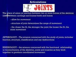
Articulations The place of union or junction between two or more bones of the skeleton (also between cartilage and bones/teeth and bones - allow for movement - structure of joint determines the range of movement - the closer the fit, the stronger the joint; the looser the fit, the more movement ARTHROLOGY – The science concerned with the study of joints, includes function, structure, classification and any dysfunctions KINESIOLOGY – the science concerned with the functional relationship, or biomechanics, of the skeleton, joints and muscles as they work together to produce coordinated movement.
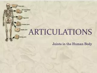
Classification of joints • A. Structural classification – based on the presence or absence of a joint cavity and the kind of supportive tissue surrounding the joint. • Three types: • 1. Fibrous joints – lack a joint cavity; fibrous connective tissue connects articulating joints • 2. Cartilaginous joints – lacks a joint cavity; cartilage binds articulating bones • 3. Synovial joints – has a joint cavity; ligaments help support articulating bones
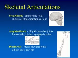
B. Functional classification – based on the degree of movement permitted within the joint • Three types: • Synarthrosis – immoveable joints • Amphiarthrosis – slightly moveable joints • Diarthrosis – freely moveable joints
II. Breakdown by functional classification • Synarthroses (immoveable joints) • 1.Suture – strongest joint by structure • - fibrous joints found between flat bones of the skull • - irregular structure that gives strength and reduces fractures • 2.Gomphoses • - fibrous joints that occur between teeth and the supporting bones of the jaw. • - located where the root of a tooth is attached to the periodontal ligament of the alveolus (socket) of the bone.
3. Synchondroses - cartilaginous joint with hyaline cartilage as the connective tissue - some are temporary and form the epiphyseal growth plates between the diaphysis and epiphysis in the long bones of children
B. Amphiarthroses – slightly moveable joint • Syndesmoses • Fibrous joint found only in the forearm and leg where adjacent bones are held together by collagenous fibers • Characteristic of the side-to-side joints between tibia-fibula and the radius-ulna (allows rotation) • 2. Symphyses (symphysis pubis, and intervertebral discs) • Cartilaginous joint separated by a pad of fibrocartilage; allows for limited movement • Only limited motion is possible at each joint, the combined movement allows for extensive movement (vertebrae)
C. Diarthroses – freely moveable • Characteristics: • a. provides a wide range of precise, smooth movements while maintaining stability, strength, and some rigidity in the body • b. most complex and varied of the three major types • c. range of movement is limited by three factors: - structure of the bones participating in the articulation - the strength and tautness of the associated ligaments, tendons and joint capsule - the size, arrangement and action of the muscles that span the joint (“double jointed” is a misnomer; not two joints, but extreme maneuverability due possibly to loose ligaments and tendons)
2. Structure of the synovial joint a. Synovial Cavity – space between the articulating bones refers to the structural classification b. Articular Cartilage – present in all diarthrotic joints (2 mm thick), hyaline cartilage covers the articulating surface c. Articular Capsule – surrounds the entire diarthrotic joint (two layers): FIBROUS LAYER – outer layer - dense, irregular connective tissue - attaches to periosteum of bones - permits movement and resists dislocation - fibers from ligaments; hold bones together
SYNOVIAL MEMBRANE – Inner layer - secretes synovial fluid (looks,feels like uncooked egg white) - lubricates joints and nourishes cartilage - houses phagocytic cells that remove microbes and debris
d. Accessory ligaments – some outside of articular capsule, some within • e. Articular discs – pads of fibrocartilage called menisci (meniscus = singular) • Stabilize joint by forming tighter fit • Tearing of these is called torn cartilage • f. Bursae – sac-like structures between moving parts (help cushion and reduce friction • Filled with synovial fluid • Found between skin and bone, tendon and bone, muscle and bone, ligaments and bone
3. Types of synovial joints • Gliding joint – simplest type • - Allow only side-to-side and back-and-forth movements with minimal rotation • Surfaces are usually flat or slightly concave/convex • EX: Intercarpal/intertarsal, sternoclavicular, between adjacent vertebrae • b. Saddle joint- looks like a saddle • Each articular process has a concave surface in one direction, convex in the other • Is a modified condyloid joint allowing a wider range of movement • EX: only associated with the thumb (located at the articulation of the trapezium of the carpus with the first metacarpal bone)
c. Hinge joint – permits bending in only one direction • (similar to the hinge of a door) • One surface is always concave and the other is convex • Most common type of synovial joint • EX: knee, humeroulnar, phalanges
d. Pivot – movement is limited to rotation about a central axis • One surface is rounded and fits into a depression of another • EX: proximal articulation of the radius and ulna, articulation between atlas and axis • e. Ball-and socket – formed by articulation of a rounded convex surface with a cuplike cavity • Provides the greatest range of movement of all joints • EX: hip and shoulder joints f. Condyloid or ellipsoid – structured so that an oval, convex surface of one bone fits into an elliptical, concave depression of another bone - Allows for angular movement in two directions (up and down and side-to-side motion) - EX: radiocarpal joint
III. Problems • Clinical considerations • 1. Hyperextension • 2. Strained joint • 3. Sprain • 4. Luxation • 5. Bursitis • 6. Tendonitis B. Diseases of joints Arthritis - 1. Rheumatoid arthritis 2. Osteoarthritis 3. Gouty arthritis
www.crnasomeday.com/anatpages/joints.htm www.brazoria-county.com/sheriff/images/jpg/id... www.mnsu.edu/.../humananatomy/images/body.jpg healthcare.utah.edu/healthinfo/images/ei_0276.gif commons.bcit.ca/.../pics/symphysis.jpg cache.eb.com/eb/image?id=72183&rendTypeId=35 images.main.uab.edu/healthsys/ei_0244.gif content.answers.com/.../dental/f0475-01.jpg www.hawaii.edu/.../pediatrics/pemxray/v1c18f.jpg academic.wsc.edu/faculty/jatodd1/351/joints2.jpe www.daviddarling.info/images/synovial_joint.jpg cache.eb.com/eb/image?id=72183&rendTypeId=35 sciencefun4all.net/.../Images/Joints/GLIDING.jpg www.shockfamily.net/skeleton/SADDLE.JPG www.mc.edu/.../carastafford2_files/image010.jpg sciencefun4all.net/.../Images/Joints/HINGE.jpg