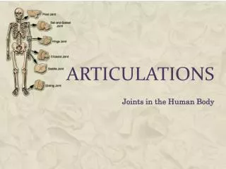
Understanding the Types of Joints: Fibrous, Cartilaginous, and Synovial
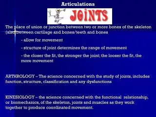
This overview explores the various types of joints in the human body, categorized into fibrous, cartilaginous, and synovial joints. Fibrous joints, which include sutures and syndesmoses, exhibit little to no movement and are characterized by tightly held bone ends through fibrous connective tissue. Cartilaginous joints allow slight movement and involve bones united by cartilage. Synovial joints, the most common type, enable free movement and possess a fluid-filled cavity and several essential anatomical features. Learn about their structures, functions, and examples to better understand joint mechanics.

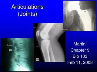
Understanding the Types of Joints: Fibrous, Cartilaginous, and Synovial
E N D
Presentation Transcript
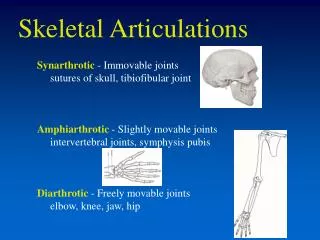
Fibrous Joints • Include all articulations where bones are held tightly together by fibrous connective tissue—called sutural ligament • Very little material separates the ends of bones, no appreciable movement is allowed • Also called synarthroses (syn=together, arthron=joint)—nonmovable joint • Two Types: • Sutures • Syndesmoses
Sutures • Bone interdigitations (grooves) fit closely and firmly together • Connecting fibers spanning between bones very short • Found only in flat bones of skull • In early adulthood, fibers of suture are replaced by bone • Bones fuse together • Called synostosis (held together by bone)
Syndesmoses • Syndesmosis=together by bands • Held together by fibrous connective tissue • Bone ends farther apart than in suture, thus connective tissue fibers joining bones longer • Bones not held as firmly as suture, thus a “give” motion occurs • Examples: • Joint between distal ends of tibia and fibula • Joints in the mid-radius/ulna and the mid-tibia/fibula
Cartilaginous Joints • Bones united by cartilage • Sight movement is possible—called amphiarthroses (amphi=on both sides) • Two types • Synchondroses • symphyses
Synchondroses • “together by cartilage” • Held together by hyaline cartilage • Many temporary, eventually replaced by bone • Examples • Area between the epiphyses and the diaphysis of long bones • Certain skull bones • Joint of first ten ribs and the costal cartilage is a permanent synchondroses
Symphyses • Aricular surfaces covered with thin layer of hyaline cartilage • A fibrocartilaginous pad separates the bones—the pads (discs) are compressible, serve as shock absorbers • Examples: • Intervertebral discs: pads between adjacent vertebrae • The junction of the pubis bones—pubic synapse • The midline symphysis between the two halves of the mandible
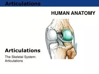
Synovial Joints • The majority of joints • Freely movable—limited by ligaments, muscles, tendons, or adjoining bones • Referred to as diarthroses=“through joint” indicating only slight limitations to the movement of such joints • Have a fluid-filled joint cavity • Four distinguishing features: • Articular cartilage (thin layer of hyaline cartilage covering surface of bone • Articular capsule (a double-layer membrane surrounding joint) • Synovial membrane (loose connective tissue whose inner surface supplied by capillaries • Synovial fluid (thick fluid that provides nutrients to articular cartilages, lubricates joint)
Synovial Joints continued • Synovial joint also may have articular discs or menisci • Articular discs divide synovial cavity into two separate cavities • Examples: • Jaw • Sternoclavicular joint • Distal radioulnar joint • Synovial membranes form two structures that are not actually part of the synovial joint but are associated with them: • Bursae: a small sac lined with synovial membranes. Act as cushions between the structures they separate • Some are subcutaneous—between bone and skin • Most located between tendons and bone • Tendon Sheaths: cylindrical synovial sacs that wrap around tendons, found where tendons cross joints
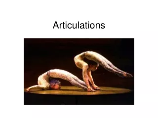
Synovial Joint Movement • Identified by the type of movement they permit • Many have axis of rotation • Uniaxial joints—have only one axis of motion and move on one plane (elbow and knee) • Biaxial joints—have two axes allowing movement in two planes that are at right angles to each other (between tarsals and metatarsals, carpals and metacarpals) • Triaxial joints—more than two axes and move in three planes (hip and shoulder) • General movements: gliding, angular, circumduction, and rotation
Gliding • Simplest most common type of movement • Surfaces of adjoining bones move back and forth • The articulating surfaces are flat or slightly concave • Examples: • Joint between head of ribs and the bodies of the vertebrae • Joint between the tubercles of the ribs and the transverse processes of the vertebrae • Intercarpal and intertarsal joints
Angular Movement • Flexion • When a bone is moved in an anterior-posterior plane in a manner as to decrease the angle between in and the adjoining bones • Example: bending the elbow, bringing the thigh up toward the abdomen, bringing the calf of the leg up toward the back of the thigh • Pulling the heel upward, lowering the toe region of the foot is plantar flexion • Extension • Opposite of flexion • Causes the angle between adjoining bones to increase • Occurs when a flexed joint is placed back in its anatomical position • Hyperextension occurs when part is moved beyond a straight position—arching the back or bringing the limbs posteriorly beyond the plane of the body • Raising the toe region toward the shin is called dorsiflexion
Angular movement continued • Abduction • When a part is moved away from the midline of the body • Example, moving the fingers away from the midline of the hand • Adduction • The opposite of abduction • When a part is moved toward the midline of the body • Example: moving the fingers toward the midline of the hand • Circumduction • Delineates a cone, the base of the bone is outlined by the movement of the distal end of the bone, with the apex of the cone lying in the articular cavity • Movement combines flexion, abduction, extension, and adduction • Example: shoulder and hip
Angular movement continued • Rotation • The motion of a bone around a central axis without any displacement of that axis • If the anterior surface of a bone moves inward, called medial rotation • If the anterior surface turns outward it is lateral rotation • Supination • The outward rotation of the fore-arm, causing the palm to face upward or forward and the radius and ulna to be parallel • Supination occurs when the arms in the anatomical position • Pronation • The inward rotation of the fore-arm, causing the radius to cross diagonally over the ulna and the palm to face downward or backward
Angular Movement continued • Special movements • Elevation: raising the part, examples: raising the scapula or mandible • Depression: lowering the part, example: lowering scapula or mandible • Inversion: twisting of the foot so that the sole faces inward with its inner margin raised • Eversion: twisting of the foot so that the sole faces outward with its outer margin raised • Protraction: the motion that moves a part forward, such as the mandible • Retraction: the motion that returns a protracted part to its usual position
Types of Synovial Joints • Nonaxial joints • Gliding (arthrodial) joints: formed primarily by the apposition of flat, or only slightly curved, articular surfaces. Movement is allowed in any direction, being llimited only by ligaments or bony processes that surround the articulation. Example: vertebrae and between most carpal and tarsal bones • Uniaxial joints • Hinge (ginglymus) joints: the articular surfaces are shaped such that the only movements possible are flexion and extension. Examples: elbow, knee, and joints between the phalanges • Pivot (trochoid) joints: rotation around the longitudinal axis of the bone. Examples: rotation of atlas around the axis, proximal articulations between the radius and ulna
Types of Synovial joints continued • Biaxial Joints • Condyloid (ellipsoid) joints: have one articular surface slightly concave and other slightly convex, movement is allowed in two planes that are at right angles to each other. Flexion, extension, abduction, and adduction can occur at condyloid joints. Circumduction is possible, but axial rotation is not. Examples: between radius and carpals, the occipital condyles of skill and the atlas, metacarpophalangeal and metatrasophalangeal joints • Saddle joints: allow the same movement as the condyloid joints. The articular surface of each bone is concave in one direction and convex in the other; the bones fit together just as two saddles would if the riding surface of one were rotated 90 degrees. Only one saddle joint, the thumb • Triaxial Joints • Spheroid (ball and socket) joints: formed by a spherical head of one bone fitting into a cup-shaped cavity on another. Movement in indefinite number of axes—flexion, extension, abduction, adduction, circumduction, medial and lateral rotation. Only 2—shoulder and hip
Clinical Significance of Articulations • Sprains • Result from the twisting or overstretching of a joint causing a ligament to tear or separate from bony attachment---excessive tissue fluid accumulates and causes swelling • Dislocations • Articular surfaces are forcibly displaced • Severe dislocations result in bones and surrounding tendons and ligaments being damaged • Most common in shoulder, thumb, and fingers
Clinical Continued • Bursitis • When one or more bursae surrounding a joint becomes inflamed • May result from injury, heavy exercise, or infection • Results in discomfort and limiting motion • Tendinitis • The inflammation of tendon sheaths around a joint • Local tenderness at the point of inflammation and severe pain upon movement of joint • Results from trauma to, or excessive use of a joint • Most common in wrist, elbow, or shoulder
Clinical continued • Slipped Disc • The relatively soft nucleus pulposa within the intervertebral disc is squeezed to the side of the disc • Results from trauma or improper distribution of weight along the vertebral column resulting in poor posture • Causes the disc to protrude and/or rupture—result in severe pain along the path of nerves, numbness, if nerve damage results—weakness and degeneration of muscles
Clinical continued • Torn Menisci • Sudden changes of direction while bearing body weight can cause the menisci of knee to tear loose • Causes severe pain and swelling of the joint • Arthroscopic surgery repairs damaged menisci • Arthroscope (needlelike viewing instrument ) inserted in the knee • Fiber-optic light allows doc to see injury and small incisions are made to insert cutting instruments • Performed under local anesthesia, patient returns home same day
Clinical Continued • Arthritis • Pathological changes in the joint membranes, cartilage, and bone cause swelling and pain • Causes of arthritis unknown, but trauma, infection by bacteria (staphylococci, streptococci, and gonococci) and metabolic disorders have been implicated • May be genetically inherited • Types: • Osteoarthritis • Rheumatoid arthritis • Gouty arthritis
Osteoarthritis • Most common form of arthritis • Chronic inflammation that causes the articular cartilage in the affected joint to degenerate • Causes pain, swelling, and stiffness • As articular cartilage degenerates, bony spurs develop and restrict the movement of the joint
Rheumatoid Arthritis • Severely damages the joint • Affects principally small joints of the body—hands, feet, knees, ankles, elbows, and wrists • Affects women more than men • Begins with inflammation of the synovial membrane of the joint, swelling and pain occur--Inflamed synovial fluid produces pannus (abnormal tissue that grows over surface of articular cartilage) • Articular cartilage beneath pannus is destroyed, pannus fills joint space and becomes invaded by fibrous tissue—restricts movement • Calcification of pannus may ankylose (fuse) the joint
Gouty Arthritis • Gout is characterized by sudden severe pain and swelling of joint • Primarily affects toes, insteps, ankles, heels, knees, and wrists • More common in males • Inherited genetic defect that causes either and increased production of uric acid or a reduction in the ability of the kidney to excrete uric acid • Causes hypouricemia (increase level of uric acid in blood) • Body fluids become supersaturated and result in the formation of sodium urate crystals in the soft tissues of the body as well as joints • Causes inflammation that eventually may erode the articular cartilage and underlying bone—causes intense pain and immobility
Effects of Aging • Aging causes a progressive loss of cartilage surface of joints • Called degenerative osteoarthropathy • Varies greatly from individual to individual • Women develop bony swellings (Heberden’s nodes) in terminal phalanges • Men most commonly develop degenerative osteoarthropathy n spine • By age 80, virtually everyone has some degenerative osteoarthropathy of knee and elbow, and to some degree hip and shoulder • Chronic pain and pressure result