Download

1 / 119
1.2k likes | 1.44k Vues
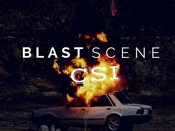
BLASTS AND BURNS: Don’t Feel The Heat!. Susan Marie Baro , DO, FACOS Associate Trauma and Surgical Critical Care Associate Director Surgical Critical Care Physician Director Blood Conservation Program. OBJECTIVES.

E N D
BLASTS AND BURNS: Don’t Feel The Heat! Susan Marie Baro, DO, FACOS Associate Trauma and Surgical Critical Care Associate Director Surgical Critical Care Physician Director Blood Conservation Program
OBJECTIVES • Understand the injuries that result from explosions and review current management and treatment of Blast Injuries • Review Burn Injury Classifications and Standard Treatments • Calculate % TBSA in Burns • Calculate IV Fluid Requirements in Burns
AMERICAN BURN ASSOCIATIONBurn Injury Severity Grading System • Minor Burn • 15% TBSA (Total Body Surface Area) or less in adults • 10% TBSA or less in children and the elderly • 2% TBSA or less full thickness burn in children or adults without cosmetic or functional risk to eyes, ears, face, hands, feet or perineum
AMERICAN BURN ASSOCIATIONBurn Injury Severity Grading System • Moderate Burn • 15 – 25% TBSA in adults with less than 10% full thickness burn • 10 – 20% TBSA partial thickness burn in children < 10 and adults > 40 years of age with less than 10% full thickness burn • 10% TBSA or less full thickness burn in children or adults without cosmetic or functional risk to eyes, ears, face, hands, feet, or perineum
AMERICAN BURN ASSOCIATIONBurn Injury Severity Grading System • Major Burn • 25% TBSA or greater • 20% TBSA in children <10 and adults > 40 years of age • 10% TBSA or greater full thickness burn • All burns involving eyes, ears, face, hands, feet, or perineum that are likely to result in cosmetic or functional impairment
AMERICAN BURN ASSOCIATIONBurn Injury Severity Grading System • Major Burn (cont.) • All high voltage electrical burns • All burn injury complicated b y major trauma or inhalation injury • All poor risk patients with burn injury
CLASSIFICATION OF BURNS • Thermal • Cold Exposure • Chemical • Electrical Current • Inhalation • Radiation
CLASSIFICATION BASED ON DEPTH OF TISSUE INJURY • 1st Degree – Superficial or Epidermal • 2nd Degree – Partial Thickness • 3rd Degree – Full Thickness • 4th Degree – burns extending beneath the subcutaneous tissues involving the fascia, muscle, and /or the bone
SUPERFICIAL BURN • Epidermal layer (ex, sunburn) • No Blisters • Red, painful, and dry • Epidermal layer peels away • Blanches with pressure • Subsides over 2 – 3 days and heals within 6 days without scarring
PARTIAL THICKNESS: SUPERFICIAL • Between the epidermis and the dermis • Forms blisters within 24 hours • Painful, red, weeping • Blanches with pressure • Pigment changes can occur • Usually heals in 7 – 21 days • Scarring unusual
PARTIAL THICKNESS:DEEP • Extends deep into the dermis • Damages hair follicles and glandular tissue • Painful to pressure only • Almost always blisters • Wet, waxy, or dry • Variable mottled coloration (Patchy cheezy white to red)
PARTIAL THICKNESS:DEEP (cont). • Does not blanch • Heals in 3 – 9 weeks if no grafting required • Causes hypertrophic scarring • If involves the joint, expect dysfunction even with aggressive physical therapy • Hard to differentiate from Full Thickness burn
FULL THICKNESS • Extends through and destroys all layers of the dermis and often injures underlying subcutaneous tissue • Burn eschar and denature dermis usually intact • Eschar compromises viability of limb and torso if circumferential • Anesthetic or hypoesthetic
FULL THICKNESS (cont.) • Skin waxy white to leathery gray to charred and black • Dry and inelastic • Does not blanch • No vesicles or blisters
FULL THICKNESS (cont.) • Eschar usually separates from the underlying tissue and reveals an unhealed bed of granulation tissue • Without surgery – they heal by wound contracture with epithelialization around the edges • Scarring is severe with contractures
FOURTH DEGREE • Deep • Potentially life threatening • Extend through the skin to underlying structures
TOTAL BODY SURFACE AREA • Size is usually underestimated • Results in under resuscitation • Lund-Browder • Most accurate for both children and adults • Takes into account the relative % of body surface area affected by growth • Kids have larger heads and smaller extremities
TOTAL BODY SURFACE AREA (cont). • Rule of Nines (adults) • Each leg represents 18% TBSA • Each arm represent 9% TBSA • Anterior and Posterior Trunk each represent 18% TBSA • Head represents 9 % TBSA
TOTAL BODY SURFACE AREA (cont). • Palm Method • Used when the burn is irregular and/or patchy • Utilizes the surface area of the patients palm • Palm, excluding extended fingers = 0.5% patients TBSA • Palm, extending fingers = 1% of patients TBSA
INITIAL MANAGEMENT • Essentially ATLS • Special attention to respiratory distress and smoke inhalation • Remove clothing promptly • Consider early transfer to Burn Center • History is important • Materials, chemicals, open vs closed space, explosion or blast involvement, associated trauma
AIRWAY • Inhalation injury remains a leading cause of death in the adult burn victim • Present in 2/3’s of patient with burns > 70% TBSA • Supplemental oxygen, maintain airway • Upper airway edema occurs rapidly
AIRWAY (cont.) • RSI with Succinylcholine acceptable in the first 72 hours but no later secondary to the risk of severe hyperkalemia • Significant % develop ARDS
SIGNS OF SIGNIFICANT SMOKE INHALATION INJURY • Persistent cough, stridor, or wheezing • Hoarseness • Deep facial or circumferential neck burns • Nares with inflammation or singed hair • Carbonaceous sputum or burnt matter in the nose or mouth • Blistering or edema of the oropharynx
SIGNS OF SIGNIFICANT SMOKE INHALATION INJURY (cont.) • Depressed mental status • Respiratory distress • Hypoxia or Hypercapnia • Elevated Carbon Monoxide and/or Cyanide levels • Inhalation injury from hot gasses usually occurs above the vocal cords
CARBON MONOXIDE AND CYANIDE • Check Carboxyhemaglobin level in all patients with moderate to severe burns • Standard Pulse-Ox not reliable • Treatment with high flow oxygen alone effectively removes CO • Hyperbaric Oxygen Treatment if increased CO or if treatment for Cyanide poisoning places patient at risk for hypoxemia
CARBON MONOXIDE AND CYANIDE (cont.) • Check Methemaglobin if Cyanide poisoning suspected • Consider Cyanide toxicity in severe burn patients with unexplained lactic acidosis and declining EtCO2 • Treatment: Hydroxocobalamin
TREATMENT • Supplemental Oxygen and Airway Protection • Bronchodilators when bronchospasm present • Avoid Corticosteroids • Fluid resuscitation with aggressive monitoring
TREATMENT (cont.) • Vent Settings: low tidal volumes to minimize airway pressures and to reduce incidents of Ventilator Associated Acute Lung Injury (ALI) • Inhaled Nitric Oxide – may increase hypoxic vasoconstriction • Aerosolized Heparin and N-Acetylcysteine(NAC) – may help to remove broncho-pulmonary casts
FLUID RESUSCITATION • Burn Shock – occurs within 24 – 48 hours • Characterized by myocardial depression and increased capillary permeability • Results in large fluid shifts and depletion of intravascular volume • Rapid, aggressive fluid resuscitation helps to reconstitute the intravascular volume and maintain end organ perfusion
FLUID RESUSCITATION (cont.) • A-line • Foley for accurate urine outputs • Over-resuscitation leads to ARDS, pneumonia, MSOF, and compartment syndromes (including abdomen, limb, and orbit) • Any patient with > 15% TBSA, nonsuperficial burns (2nd/3rd Degree) should receive formal fluid resuscitation
FLUIDS • IV Crystalloid – typically Ringer’s Lactate • helps to reduce incidence of hyperchloremic acidosis associated with large volumes of isotonic saline (NS) • Colloid and Hypertonic Saline for initial resuscitation not found to show any improvement in outcomes, are more expensive, and possibly increase renal failure and death
FLUIDS (cont.) • Following initial resuscitation IV fluids need to meet baseline fluid needs and maintain Urine outputs • IF UO < 0.5 ml/kg/hr – bolus with 500 to 1000 ml fluid and increase rate by 20 – 30% • If adequate resuscitation and patient stabilizes, change to D5 ½ NS with 20 mEqKCl per liter at maintenance to keep UO > 0.5 ml/kg/hr
ESTIMATING INITIAL FLUID REQUIREMENTS • Parkland Formula – utilized in initial 24 hrs • Includes partial and full thickness burns • 4 ml/kg for each % of TBSA burned over 15% TBSA • ½ volume given in 1st 8 hours and the remaining volume given over the next 16 hours
ESTIMATING INITIAL FLUID REQUIREMENTS (cont.) • Modified Brooke Formula • Given over initial 24 hours • 2 ml/kg for each % TBSA • Likely reduces the overall volume
BLOOD TRASFUSION • Avoid if possible • Associated with increased mortality • Only if Hemoglobin < 8 gm/dL unless patient with acute coronary syndrome • If at risk for ACS – transfuse to 10 gm/dL
IMMEDIATE BURN CARE • Remove clothing • Cool burned area immediately using cool water or saline soaked gauze • can minimize the zone of injury in small burns • Monitor cor body temp to prevent hypothermia, especially if >10% TBSA • Avoid temps below 35o C/95o F • Aggressive Pain control with Morphine and Benzo’s for anxiety
CHEMOPROPHYLAXIS • Extensive burns cause immunosuppression on basis of altered neutrophil activity, T lymphocyte dysfunction, and imbalance in production of cytokines • Bacterial colonization of the burn eschar site can result • Burns destroy physical barrier to tissue invasion • Permits spread of bacteria to the dermis and through the lymphatics along the fibrous septae
CHEMOPROPHYLAXIS (cont.) • Once invasion occurs – organisms can invade the blood vessels producing secondary bacteremia • Topical antibiotics are given to all patients with nonsuperficial burns
TETANUS • Update for any burns deeper than superficial • Tetanus Immune Globulin – if patient did not receive complete set of primary immunizations