Therapeutic Ultrasound
340 likes | 857 Vues
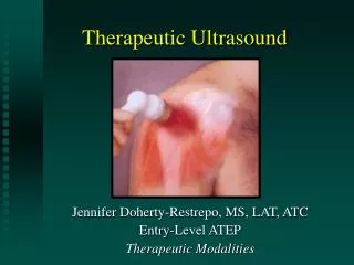
Therapeutic Ultrasound. Clinical Application. FDA Labeling Requirements. Output frequency Effective Radiating Area (ERA) Beam Nonuniformity Ratio (BNR) Beam profile Date of last service/calibration should also be posted on the unit. Treatment Area. US heats a limited area

Therapeutic Ultrasound
E N D
Presentation Transcript
Therapeutic Ultrasound Clinical Application
FDA Labeling Requirements • Output frequency • Effective Radiating Area (ERA) • Beam Nonuniformity Ratio (BNR) • Beam profile • Date of last service/calibration should also be posted on the unit
Treatment Area • US heats a limited area • About the size of a catsup packet • Treatment area should be 2 to 3 times the size of the ERA • For larger areas, divide the treatment area into smaller zones
Coupling Methods Direct CouplingImmersion MethodPad/Bladder Method
Coupling Methods • Ultrasonic energy cannot pass through the air • A coupling medium is required • Medium should be water-based • Coupling method should confirm to the body area • The body area should be clean and relatively hair-free
Direct Coupling • Gel or Creams • Only use approved coupling agents • Apply liberally to area • Remove air bubbles by passing sound head over area (before power is increased)
Direct Coupling • Move the sound head s-l-o-w-l-y • 4 cm/sec • Moving the head faster decreases heating • If the patient describes discomfort, decrease the output intensity
Coupling Ability of Various Media Substance Transmission • Saran Wrap 98 • Lidex ge, fluocinonide (.05%) 97 • Thera-Gesic 97 • Mineral oil 97 • US Transmission gel 96 • US Transmission lotion 90 • Chempad-L 68 • Hydrocortisone powder (1%) 29 • Hydrocortisone powder (10%) 7 • Eucerin cream 0 • Myoflex 0 • White petrolatum gel 0
Immersion Technique • Used to treat irregularly shaped areas • The limb is immersed in a tub of degassed water • If tap water is used, increase the output intensity by 0.5 w/cm2 • Transducer is held appx. 1” from the body part • Avoid the formation of air bubbles
Pad (Bladder) Method • A mass of conductive gel • Commercial pads • Self-made bladders • Conforms to the treatment area • Commercial pads help limit the size of the treatment area
Output Parameters Output FrequencyDuration Duty CycleOutput Intensity
Output Frequency • Determines the treatment depth • 1 MHz Output • Deep (5 to 7 cm) tissues • Rotator cuff, vastus intermedius, gastroc • 3 MHz Output • Superficial (up to 3cm deep) tissues • Patellar tendon, MCL, brachialis • Remember that adipose tissue is transparent to ultrasound
Treatment Duration • Depends on: • Size of the treatment area • Output intensity • Therapeutic goals • Vigorous heating • 1 MHz output • 10 to 12 minuts • 3 MHz output • 3 to 4 minutes • Also see “Output Intensity”
Duty Cycle • Determine the proportion of thermal and nonthermal effects • High duty cycle: Predominantly thermal effects • Low duty cycle: Predominantly nonthermal effects • Thermal effect used in subacute and chronic conditions • Nonthermal effects may be beneficial in acute stages
Output Intensity – 1 MHz • At a 100% duty cycle • 1.75 W/cm2 output • Approximately 4 min treatment is required to increase tissue temp 4°C
Output Intensity – 3 MHz • At a 100% duty cycle • 1.5 W/cm2 output • Approximately 10 min treatment is required to increase tissue temp 4°C
Ultrasound and Electrical Stimulation “Combo Treatment”
US/Electrical Stimulation • Theoretically combines the benefit of the two treatments • Used to treat: • Trigger points • Muscle spasm • Output parameters: • Thermal ultrasound • Motor level electrical stimulation
Acute injuries (100% duty cycle) Ischemic areas Areas of impaired circulation including arterial disease Over areas of deep vein thrombosis Anesthetic areas Over cancerous tumors Over sites of active infection or sepsis Over the spinal cord or large nerve plexus in high doses Exposed metal that penetrates the skin (e.g., external fixation devices) Areas around the eyes, heart, skull, or genitals Over the thorax in the presence of an implanted pacemaker Pregnancy when used over the pelvic or lumbar areas Over a fracture site before healing is complete Stress fracture sites or sites of osteoporosis Over the pelvic or lumbar area in menstruating female patients Contraindications
Precautions • Symptoms may increase after the initial treatments. • Use caution when applying ultrasound around the spinal cord, especially after laminectomy. • The use of ultrasound over metal implants is not contraindicated • Keep the sound head moving • Use caution when applying ultrasound over epiphyseal plates of growing bone
Instrumentation • Duty cycle: Adjusts between continuous and pulsed ultrasound application • Frequency: Selects the depth of penetration • Gel warmer: Used to preheat the transmission gel, primarily for patient comfort • Intensity: Adjusts the output intensity. • The WATT METER displays the output in either total watts or watts per square centimeter. • Maximum head temperature: Prevents overheating in case the head is not properly coupled. • Pause: Interrupts the treatment but retains the remaining amount of treatment time • Power: Turns the unit ON or OFF • Start-Stop: Initiates or terminates the production of ultrasound from the transducer. • Timer: Sets the duration of the treatment. • Watt meter: Displays the output of ultrasound in total watts or watts per square centimeter.
Patient Preparation • Establish that no contraindications are present • Determine the method and mode of ultrasound application to be used • Clean the area to be treated • Determine the type of coupling method to be used • Identify a treatment area that is 2 to 3 times the ERA • For direct coupling: spread the gel over the area to be treated • Explain the sensations to be expected during the treatment: • Mild to moderate warmth (but not pain or burning) for thermal treatments • No subcutaneous sensations during pulsed ultrasound • For thermal treatments pre-heating with a moist heat pack will decrease the treatment time required to reach vigorous heating levels • Advise the patient to report any adverse, unusual, or painful sensations during the treatment.
Initiation of the Treatment • Reduce the INTENSITY to zero before turning on the POWER. • Select the appropriate mode for the output (CONTINUOUS or PULSED) • Set the WATT METER to displays the appropriate output for the type of treatment • Set the TIMER to the appropriate treatment duration • Begin slowly moving the sound head over the medium • Press the START button to begin the treatment session • Units having low BNR may be moved at a slower rate than those with a higher BNR • Slowly increase the INTENSITY • Keep the sound head moving • Move the head at a moderate pace (4 cm per second or slower) • Use firm, yet not strong, overlapping strokes • If pain is experienced: • Move the sound head at a faster rate • Use a lower duty cycle • Use a lower the intensity • If the gel begins to wear away or if the sound head begins sticking on the skin, depress the PAUSE button and apply more gel
Phonophoresis Application • Preheating the area to increase the absorption of the medication • Use only approved ultrasound transmission media. • The direct coupling method is recommended • Efficacy of phonophoresis using the bladder method has not been established • Ensure that the skin is well moistened; avoid areas of dry skin • Position the extremity to encourage circulation. • Use a continuous output to maximize the effect of phonophoresis • Unless the thermal effects of ultrasound are contraindicated • Follow the procedures described in “Initiation of the Treatment” • After treatment, cover the remaining medication with an occlusive dressing.
Termination of the Treatment • If the treatment is being terminated prematurely: • Reduce the intensity before removing the transducer • Clean the remaining gel from the patient’s skin • To ensure continuity of treatment sessions: • Record the treatment parameters • Output frequency • Intensity • Duration • Duty cycle • Running count of ultrasound treatments given for this condition • Immediately initiate any post-treatment stretching
Maintenance Daily Maintenance • Clean ultrasound head and transducer face as recommended by the manufacturer Monthly Maintenance • Check all electrical cords for frays or kinks • Check the sound head cable for frays or kinks • Clean the transmitter face as recommended by the manufacturer. Federal regulations require that therapeutic ultrasound units be recalibrated annually by an authorized service technician