Figure 27.1
Ureter. Urinary bladder. Prostatic urethra . Peritoneum. Membranous urethra . Seminal vesicle . Ampulla of ductus deferens. Urogenital diaphragm . Pubis. Ejaculatory duct . Corpus cavernosum . Corpus spongiosum . Rectum. Prostate. Spongy urethra. Bulbourethral gland .
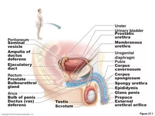

Figure 27.1
E N D
Presentation Transcript
Ureter Urinary bladder Prostatic urethra Peritoneum Membranous urethra Seminal vesicle Ampulla of ductus deferens Urogenital diaphragm Pubis Ejaculatory duct Corpus cavernosum Corpus spongiosum Rectum Prostate Spongy urethra Bulbourethral gland Epididymis Glans penis Anus Prepuce Bulb of penis Testis Ductus (vas) deferens External urethral orifice Scrotum Figure 27.1
The Scrotum • Temp kept constant by 2 muscles • Dartoswrinkles scrotal skin • Cremasterelevate testes
Urinary bladder Superficial inguinal ring (end of inguinal canal) Testicular artery Ductus (vas) deferens Spermatic cord Penis Autonomic nerve fibers Middle septum of scrotum Pampiniform venous plexus Cremaster muscle Epididymis External spermatic fascia Tunica vaginalis (from peritoneum) Superficial fascia containing dartos muscle Tunica albuginea of testis Scrotum Internal spermatic fascia Skin Figure 27.2
The Testes • Surrounded by: • Tunica vaginalis, from peritoneum • Tunica albuginea, fibrous capsule
Spermatic cord Blood vessels and nerves Ductus (vas) deferens Testis Head of epididymis Seminiferous tubule Efferent ductule Rete testis Lobule Straight tubule Septum Tunica albuginea Body of epididymis Tunica vaginalis Duct of epididymis Cavity of tunica vaginalis Tail of epididymis (a) Figure 27.3a
The Testes • Interstitial (Leydig) cells outside seminiferous tubules produce androgens
Seminiferous tubule (c) Interstitial cells Spermatogenic cells in tubule epithelium Areolar connective tissue Myoid cells Sperm Figure 27.3c
The Penis • Penis consists of • Root and shaft ends in glans penis • Prepuce, or foreskin—the cuff of loose skin covering the glans • Circumcision - surgical removal of foreskin • Crura • Proximal end surrounded by ischiocavernosus muscle; anchors penis to pubic arch
Ureter Ampulla of ductus deferens Seminal vesicle Urinary bladder Ejaculatory duct Prostate Prostatic urethra Bulbourethral gland and duct Orifices of prostatic ducts Membranous urethra Urogenital diaphragm Bulb of penis Root of penis Crus of penis Bulbourethral duct opening Ductus deferens Corpora cavernosa Epididymis Corpus spongiosum Shaft (body) of penis Testis Section of (b) Spongy urethra Glans penis Prepuce (foreskin) (a) External urethral orifice Dorsal vessels and nerves Corpora cavernosa Urethra Skin Tunica albuginea of erectile bodies Deep arteries Corpus spongiosum (b) Figure 27.4
Accessory Glands: Seminal Vesicles • Produces viscous alkaline seminal fluid • Fructose, ascorbic acid, coagulating enzyme (vesiculase), and prostaglandins • 70% of the volume of semen
Accessory Glands: Prostate • Encircles urethra inferior to bladder • Secretes milky, slightly acid fluid: • Contains citrate, enzymes, and prostate-specific antigen (PSA) • Activation of sperm
Accessory Glands: Bulbourethral Glands (Cowper’s Glands) • Pea-sized glands • Prior to ejaculation, produce thick, clear mucus • Lubricates glans penis • Neutralizes traces of acidic urine in urethra
Semen • Contains fructose, protects and activates sperm, and facilitates movement (relaxin) • Prostaglandins • Decrease viscosity of mucus in cervix • Stimulate reverse peristalsis in uterus • 2–5 ml of semen ejaculated, containing 20–150 million sperm/ml
Male Sexual Response • Erection: • Parasympathetic reflex promotes release of nitric oxide (NO) • NO causes erectile tissue to fill with blood • Expansion of corpora cavernosa • Compresses drainage veins and maintains engorgement • Corpus spongiosum keeps the urethra open
Ureter Ampulla of ductus deferens Seminal vesicle Urinary bladder Ejaculatory duct Prostate Prostatic urethra Bulbourethral gland and duct Orifices of prostatic ducts Membranous urethra Urogenital diaphragm Bulb of penis Root of penis Crus of penis Bulbourethral duct opening Ductus deferens Corpora cavernosa Epididymis Corpus spongiosum Shaft (body) of penis Testis Section of (b) Spongy urethra Glans penis Prepuce (foreskin) (a) External urethral orifice Dorsal vessels and nerves Corpora cavernosa Urethra Skin Tunica albuginea of erectile bodies Deep arteries Corpus spongiosum (b) Figure 27.4
Male Sexual Response • Ejaculation • Sympathetic reflex • Ducts and accessory glands contract and empty their contents • Bladder sphincter muscle constrict,
Meiosis • Gamete formation involves meiosis • Nuclear division in 2nto n • Produces four daughter cells • Introduces genetic variation
Mother cell (before chromosome replication) Chromosome replication Chromosome replication 2n = 4 MITOSIS MEIOSIS Tetrad formed by synapsis of replicated homologous chromosomes Replicated chromosome Prophase Prophase I Chromosomes align at the metaphase plate Tetrads align at the metaphase plate Metaphase I Metaphase Sister chromatids separate during anaphase Homologous chromosomes separate but sister chromatids remain together during anaphase I Daughter cells of mitosis Daughter cells of meiosis I No further chromosomal replication; sister chromatids separate during anaphase II 2n 2n Meiosis II n n n n Daughter cells of meiosis II (usually gametes) Figure 27.5 (1 of 2)
MITOSIS MEIOSIS Number of divisions One, consisting of prophase, metaphase, anaphase, and telophase. Two, each consisting of prophase, metaphase, anaphase, and telophase. DNA replication does not occur between the two nuclear divisions. Synapsis of homologous chromosomes Does not occur. Occurs during mitosis I; tetrads formed, allowing crossovers. Daughter cell number and genetic composition Two. Each diploid (2n) cell is identical to the mother cell. Four. Each haploid (n) cell contains half as many chromosomes as the mother cell and is genetically different from the mother cell. Roles in the body For development of multicellular adult from zygote. Produces cells for growth and tissue repair. Ensures constancy of genetic makeup of all body cells. Produces cells for reproduction (gametes). Introduces genetic variability in the gametes and reduces chromosomal number by half so that when fertilization occurs, the normal diploid chromosomal number is restored (in humans, 2n = 46). Figure 27.5 (2 of 2)
Cytoplasm of adjacent sustentacular cells Spermatogonium (stem cell) Sustentacular cell nucleus Basal lamina Type A daughter cell remains at basal lamina as a stem cell Type B daughter cell Tight junction between sustentacular cells Primary spermatocyte Secondary spermatocytes Early spermatids Late spermatids Cytoplasmic bridge Lumen of seminifer- ous tubule Spermatozoa (c) A portion of the seminiferous tublule wall, showing the spermato- genic cells surrounded by sustentacular cells (colored gold) Figure 27.7c
Spermatogenesis • Begins at puberty Type A cells maintain germ cell line at basal lamina Type B cells move toward lumen and develop
Basal lamina Type A daughter cell remains at basal lamina as a stem cell Spermatogonium (stem cell) Mitosis Type B daughter cell Growth Enters meiosis I and moves to adluminal compartment Primary spermatocyte Meiosis I completed Secondary spermatocytes Meiosis II Early spermatids Late spermatids Spermatozoa (b) Events of spermatogenesis, showing the relative position of various spermatogenic cells Figure 27.7b
Sperm • Major regions • Head: genetic region; nucleus and helmetlike acrosome containing hydrolytic enzymes that enable the sperm to penetrate an egg • Midpiece: metabolic region; mitochondria • Tail: locomotor region; flagellum
Approximately 24 days Golgi apparatus Acrosomal vesicle Mitochondria Acrosome Nucleus 1 2 Centrioles Spermatid nucleus Midpiece Head Microtubules 3 (a) Flagellum Excess cytoplasm 4 Tail 5 7 6 (b) Figure 27.8a, b
Role of Sustentacular Cells • Provide nutrients • Dispose of excess cytoplasm during spermiogenesis • Tight junctions form a blood-testis barrier • Prevents sperm antigens from escaping into blood no immune system • Sperm are not formed until puberty, they are absent during immune system development, and would not be recognized as “self”
Cytoplasm of adjacent sustentacular cells Spermatogonium (stem cell) Sustentacular cell nucleus Basal lamina Type A daughter cell remains at basal lamina as a stem cell Type B daughter cell Tight junction between sustentacular cells Primary spermatocyte Secondary spermatocytes Early spermatids Late spermatids Cytoplasmic bridge Lumen of seminifer- ous tubule Spermatozoa (c) A portion of the seminiferous tublule wall, showing the spermato- genic cells surrounded by sustentacular cells (colored gold) Figure 27.7c