Download
1 / 11
110 likes | 246 Vues
Overview of recent ORNL progress in restraint free laboratory animal imaging. Bethesda May 20, 2005. Objective.

E N D
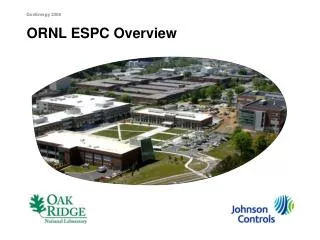
Overview of recent ORNL progress in restraint free laboratory animal imaging Bethesda May 20, 2005
Objective Briefly summarize recent ORNL activities and milestones as a part of the DOE Office of Science supported ORNL/Jefferson Lab collaboration to develop a restraint-free laboratory animal imaging system.
ORNL Participants and Roles • Justin Baba – imaging studies • Shaun Gleason – software development / motion tracking • Jim Goddard – motion tracking system • Jens Gregor (UTK collaborator) – reconstruction software • Mike Paulus – imaging system hardware, administration • Jon Wall (UTK collaborator) – animal studies
Program Objectives • Develop hardware, software and methods to correct for motion in unrestrained animal SPECT/CT studies. • Brain studies are first priority. • Approach: • MicroCT data set acquired with animal anesthetized serves as anatomic reference. • SPECT data acquired on dedicated system. • Position monitoring cameras mounted on SPECT system. • Position data used to resort SPECT events. • Reconstructed motion correct SPECT data registered with CT data.
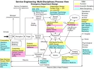
Modular System Architecture scanner motion system motion control board workstation SPECT detector system DAQ cards workstation reconstruction workstation tracking cameras video card workstation global clock
Milestones – Imaging Hardware • SPECT and CT imaging systems are now in routine use. • Improved mouse holder designed for transfer of specimens between SPECT and CT systems. • New SPECT detectors from JLAB installed and validated.
Milestones – Motion Tracking Hardware • Antireflective coating experiment for animal tube. • Investigation of polarized light for reduced reflection. • Introduction of strobed IR illumination. • Redesign of motion tracking camera mounts for improved field of view. • New reflector design adopted. • New calibration fixtures designed and implemented. • Phantom studies performed and sent to JLAB collaborators for reconstruction software development. • Live animal tracking study performed. Significant improvement in tracking observed.
Milestones – Software Development • Tracking software enhanced to support new camera geometry. • New SPECT image reconstruction package developed incorporating previously developed and new imaging reconstruction tools. • Filtered backprojection (parallel hole collimation) • MLEM (parallel hole collimation) • OSEM (parallel hole collimation) • OSEM (pinhole collimation)
Milestones – Recent Animal Studies • 18-animal SPECT/CT study comparing three new agents for imaging AA systemic amyloidosis. • 27-animal CT study to evaluate new contrast agents. • Supporting reconstruction of 300+ data sets acquired as a part of an ongoing ORNL mutant phenotyping program.
Next Steps • Convert imaging laboratory to radiation area approved for labeled live animal studies. • First motion-corrected animal studies with I-125 labeled CNS imaging compound. • Iterate with Jefferson Lab to optimize motion corrected image reconstruction. • Complete Phase I instrumentation development. • Begin fabrication of Phase II instrument for delivery to JHU collaborators. • Explore clinical opportunities.