Exploring Microscopic Worlds: Tissues and Skin Anatomy
350 likes | 431 Vues
Explore the fascinating realm of cells, tissues, and the integumentary system. Learn about the functions and classifications of epithelial and connective tissues, as well as the structure of human skin.

Exploring Microscopic Worlds: Tissues and Skin Anatomy
E N D
Presentation Transcript
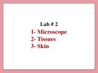
Lab # 2 1- Microscope 2- Tissues 3- Skin
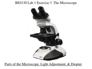
1- The Microscope Ocular lenses (10 X) Head Rotating nosepiece Objective lenses: Arm Scanning (4X) Red Low power (10X) Yellow High power (40X) Blue Mechanical Stage Oil-immersion (100X) White Condenser Light control Mechanical stage controls (X-Y knob) Diaphragm lever Fine adjustment knob Coarse adjustment knob Substage light Base
Magnification of Objective Magnification of Ocular Total Magnification Power 4 X 40 X Scanning 10 X 100 X Low 40 X 400 X High Oil-immersion 100 X 10 X 1000 X 10 X 10 X 10 X
Cells 2- Tissues Molecules Organelles Atoms Tissues The smallest particle with unique chemical identity. A particle composed of at least two atoms. Microscopic structures that carry out specific cell functions (“little organs”). They are the smallest units that carry out all the basic functions of life. A tissue is a mass of similar cells and cell products that perform a specific function. Nothing simpler than a cell is considered alive. The human body is composed of only four primary classes of tissue: Epithelial tissue Connective tissue Muscular tissue Nervous tissue
The Four Tissue Types 1- Epithelial tissue It covers exposed surfaces, lines internal passageways and chambers, and forms gland. 2- Connective tissue It fills internal spaces, provide structural support for other tissues, transport materials within the body, and stores energy reserves. 3- Muscle tissue It is specialized for contraction. - Skeletal muscle. - Cardiac muscle (the muscle of the heart). - Smooth muscle (the muscular walls of hollow organs). 4- Neural tissue It carries information from one part of the body to another in the form of electrical impulses.
Epithelial Tissue Functions of the Epithelial Tissue 1- Provide Physical Protection Epithelia protect exposed and internal surfaces from abrasion, dehydration, and destruction by chemical or biological agents. 2- Control Permeability Any substance that enters or leaves the body must cross an epithelium. The epithelial barrier can be regulated by hormones and modify by physical stress. 3- Provide Sensation Epithelia have a large sensory nerve supply that provides infor-mation about the external and internal environments. Neuroepithelia provide the sensations of smell, taste, sight, equilibrium and hearing. 4- Produce Specialized Secretions Epithelial cells that produce secretion are called glands cells.
Classification of Epithelia 1- Classification according to the number of layers Simple Stratified
2- Classification according to the cell shape 1- Squamous 2- Cuboidal 3- Columnar
1- SIMPLE squamous 2- SIMPLE cuboidal 3- SIMPLE columnar
4- STRATIFIED squamous 5- STRATIFIED cuboidal 6- STRATIFIED columnar
1- Alveoli of lungs 2- Endothelia of heart and blood vessels 1- Epidermis 2- Vagina 3- Mouth 4- Throat 5- Esophagus SIMPLE COLUMNAR 6- Rectum 7- Anus
Thyroid gland Sweat glands
Renal pelvis Ureter Urinary bladder
Stomach Intestine
Connective tissue proper Fibroblast Cartilage Connective Tissue Chondrocyte Bone Osteocyte Blood Many different types of cells 1- Cells: Each major class of connective tissue has a fundamental cell type. 2- Fibers: The fibers of connective tissue provide support. There are three types of fibers: Collagen Fibers, Elastic Fibers and Reticular Fibers. Matrix 3- Ground substance: It is the unstructured material that fills the space between the cells and contains the fibers.
1- Connective Tissue Proper Under the epithelium of organs (it forms the lamina propria of mucosa). Areolar tissue Deep to the skin, padding around eyes and kidneys. It provides padding, cushion, and insulation. It stores energy. Adipose tissue a) Loose connective tissue Reticular tissue It provides supporting and framework to organs as liver, kidneys, spleen, lymph nodes, and bone marrow. Regular Between skeletal muscles and skeleton. It forms tendons, ligaments and aponeurosis. b) Dense connective tissue Dermis, capsules of visceral organs, periosteum. It provides strength and helps to prevent overexpansion of organs such as urinary bladder. Irregular Elastic In ligaments of spinal cord and penis. It stabilizes positions of vertebrae and penis. 2- Fluid Connective Tissue a) Blood b) Lymph 3- Supporting Connective Tissue a) Cartilage b) Bone
1- Connective Tissue Proper a) Loose connective tissue AREOLAR TISSUE
b) Dense connective tissue 3- Supporting Connective Tissue a) Cartilage b) Bone
Epithelial Tissue Connective Tissue 1- Protection, transport, secretion 2- It is always in contact with an external environment 3- It is almost completely composed of cells 1- Stabilizes and supports other tissues 2- It is never in contact with an external environment 3- It is composed of cells that are separated from each other by an extracellular material called ground substance
3- Skin The Integumentary System 1- The cutaneous membrane (skin) 2- The accessory structures or appendages of the skin Nails Hair Multicellular exocrine glands
SKIN Epidermis Dermis Hypodermis 1- Epidermis and Dermis 1- Epidermis: is a keratinized, stratified squamous epithelium 2- Dermis: is a dense irregular connective tissue 3- Subcutaneous layer (Hypodermis): contains adipose tissue
Layers of the Epidermis Dead Keratinocytes Stratum Corneum Dying Keratinocytes Stratum Granulosum Langerhans cell Stratum Spinosum Keratinocytes Stratum Germinativum or Basale Merkel cell Melanocytes Basal cells (in mitosis)
2 weeks 15 to 30 days Epidermis of Soles and Palms
Subdivisions or Layers of the Epidermis 1- Stratum germinativum or basale: It is attached to the basal lamina. Its cells divide to replace superficial cells, and it contains melanocytes that produce melanin for protection against UV radiation. 2- Stratum spinosum: It is composed of 8-10 layers of cells. It contains dendritic (Langerhans) cells for immune response. 3-Stratum granulosum: It is a grainy layer whose cells produce the proteins keratin and keratoyalin. 4- Stratum lucidum: This a clear layer present only in thick skin (only in palms and soles). It contains cells filled with keratin. 5- Stratum corneum: It is composed of 15-30 layers of keratinized dead cells, and it is water resistant. It permits insensible perspiration. Functions of the Epidermis: It provides protection from chemical, physical, and biological agents.
Cells of the Epidermis 1- Keratinocytes They are the most abundant epidermal cells. Function: to produce the keratin, which is the fibrous protein that helps give the epidermis its protective properties. 2- Melanocytes They are the spider-shaped cells in the deepest layer of the epidermis. Function: To produce the pigment melanin, which protect the cell nucleus from UV light. 3- Langerhan’s cells They are start-shaped cells that ingest foreign substances and help to activate the immune system. 4- Merkel cells They are present at the epidermal-dermal junction. They are associated with a sensory nerve ending to form the Merkel disc. Function: Sensory receptor for fine, superficial touch. 5- Basal or germinative cells They are stem cells whose divisions replace the keratinocytes.
DERMIS 1- Papillary layer 2- Reticular layer 2- Dermis Functions: Thermoregulation and Protection Subdivisions or Layers of the Dermis It consists of areolar tissue (loose connective) and contains capillaries (papillary plexuses) and lymphatic vessels, as well as pain and touch receptors (Meissner’s corpuscles). It supports and nourishes the overlaying epidermis It is deep to the capillary layer. It consists of dense irregular connective tissue, which contains arteries, veins, sweat and sebaceous glands, and pressure receptors (Pacinian corpuscles). The reticular layer helps resist tension in the skin.
EPIDERMIS Dermal papillae Papillary plexus Papillary layer Tactile or Meissner’s corpuscle DERMIS Sebaceous gland Reticular layer Sweat gland Lamellated or Pacinian corpuscle Subdivisions or Layers of the Dermis DERMIS Epidermal ridges (They produce the fingerprints) (light touch receptors) (deep pressure receptors)
Cutaneous Sensation Tactile or Meissner’s corpuscle In the dermal papillae, sensible to light touch Lamellated or Pacinian corpuscle In the deep dermis or hypodermis, sensible to bumps and deep pressure Merkel disc In the deep epidermis, sensible to light touch