Tissues Chapter 3
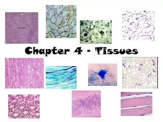
Tissues Chapter 3. Histology. Study of tissues (approximately 220 types!) Tissues: groups of highly specialized cells performing functions that benefit the organism as a whole Cells must be similar in structure & function and come from similar embryonic origin Four Primary tissue types

Tissues Chapter 3
E N D
Presentation Transcript
Tissues Chapter 3
Histology • Study of tissues (approximately 220 types!) • Tissues: groups of highly specialized cells performing functions that benefit the organism as a whole • Cells must be similar in structure & function and come from similar embryonic origin • Four Primary tissue types • Epithelium (covering) • Connective (support) • Muscle (movement) • Nervous (control) • Organs contain several tissue types, and arrangement of tissues determines organ’s structure & function
Primary Germ Layers • All tissues & organs of the body develop from one of three primary germ layers: • Ectoderm (outside) • Lining, skin, nervous • Endoderm (inside) • Organs, mucosae & glands, linings of cavities and tracts • Mesoderm (middle) • Connective tissue (i.e. blood, bone) and most muscle tissue Ectoderm Mesoderm Endoderm
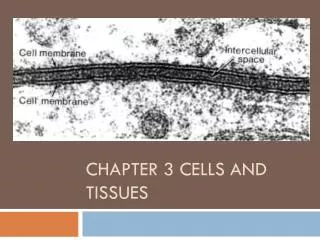
Extracellular Materials • ECF: usually fluid, but can be more gel-like or calcified (i.e. bone) • ECF provides a medium for: • Dissolving & mixing solutes • Transporting substances • Carrying out chemical reactions
Epithelial Tissue • a.k.a. epithelium (“epithe” = laid on, covering) • Lining, covering, and glandular tissue of the body • Covers all free body surfaces and contains versatile cells • Nearly all substances that the body gives off or receives must pass through epithelium • Functions: • Protection • Filtration • Absorption • Secretion
Characteristics of Epithelium • Cells fit closely together to form continuous sheets – single or multiple layers (desmosomes & tight junctions); little extracellular materials • Membranes always have one free (unattached) surface or edge (apical surface) that is exposed to body’s exterior or to the cavity of an internal organ (some have modifications like cilia or microvilli) • Basal surface (lower) of epithelium rests on a basement membrane – structureless material secreted by both epithelial cells and connective tissue cells that border the epithelium • Epithelial tissues have no blood supply of their own (avascular) and depend on diffusion from the capillaries in underlying connective tissue for food and oxygen • Regenerate easily (high mitotic rate) • Have a nerve supply • Derived from all three primary germ layers
Motion Induced Blindness Motion, your mind, and a myriad of shapes team up to rob you of your sight! Simply stare at the dot in the middle of the screen and see what happens. Don't worry, there are no lasting side effects... We think.
Classification of Epithelium • Two names: • 1st indicates relative number of cell layers • Simple (one layer) or stratified (multiple layers) or pseudostratified (looks like multiple layers) • 2nd indicates the shape of its cells • Squamous (flat) • Cuboidal (cube) • Columnar (shaped like columns) • Transitional (varies) • Stratified epithelium is named based on cells at the free surface! • Arrangement reflects location and function
Types of Epithelial Tissue • Covering & Lining Epithelium • Simple Epithelia • Simple squamous epithelium • Simple cuboidal epithelium • Simple columnar epithelium • Pseudostratified columnar epithelium • Stratified Epithelia • Stratified squamous epithelium • Stratified cuboidal epithelium • Stratified columnar epithelium • Transitional epithelium • Glandular Epithelium
Epithelial Tissue ID Quiz Tissues you need to be able to identify: • Simple squamous • Simple cuboidal • Simple columnar • Stratified squamous • Transitional • Pseudostratified Tissue/ cell parts you need to know: • Nucleus • Apical (free) surface • Basal surface • Basement membrane • Cilia • Goblet cell • Keratin
Simple Squamous Epithelium • Single layer, flat shape • All cells attached to basement membrane • Fit closely together • Forms membranes where filtration or exchange of substances by rapid diffusion occurs (absorption, secretion, filtration) • Ex: air sacs of lungs (O2 & CO2 exchanged), walls of capillaries (nutrients and gases pass between the tissue cells and blood in capillaries), form serous membranes (serosae) – slick membranes that line ventral body cavity and cover organs in that cavity • Endothelium – lines blood vessels • Mesothelium – lines body cavities and mesenteries top view JayDoc LUMEN
Simple Cuboidal Epithelium • Single layer/cube shape • Common in glands and their ducts • Ex. Salivary glands and pancreas, walls of kidney tubules, covers surface of ovaries apical surface basement membrane Tubule lining – cross-section Tubule lining – longitudinal cut
Simple Columnar Epithelium • Single layer/column (tall) shape • Often have goblet cells – produce lubricating mucus • Ex: lines entire length of digestive tract from stomach to anus • Mucosae (mucous membranes): epithelial membranes that line body cavities open to the body exterior goblet cell basement membrane (basal surface) LUMEN
Pseudostratified Columnar Epithelium • Appear to be multi-layered, but are actually one single layer that rest on the basement membrane • Nuclei appear at different heights and some cells shorter than others • Mainly functions in absorption and secretion • Can be ciliated (pseudostratified ciliated columnar epithelium) • Also can have goblet cells • Ex: respiratory tract – mucus produced by goblet cells in this epithelium traps dust and other debris, and the cilia propel the mucus upward and away from the lungs cilia cilia goblet cell LUMEN
Stratified Squamous Epithelium • Multiple layers, flat shape • Most common stratified epithelium in body • Cells at free edge are squamous, and those close to basement membrane can be cuboidal or columnar • Found in sites that receive a good deal of abuse or friction • Keratin – protein coating on apical surface (i.e. skin) • Can be keratinized or non-keratinized • Ex. Esophagus, mouth, outer portion of skin apical surface keratin basal surface Keratinized Non-keratinized LUMEN
Stratified Cuboidal Epithelium • Two cell layers with (at least) the surface cells being cuboidal in shape • Fairly rare in body; distribution extremely limited • Mainly in ducts of large glands (larger ducts of mammary glands, sweat and salivary glands, pancreas)
Stratified Columnar Epithelium • Multiple layers/Columnar cells • Basal cells vary in size and shape, see multiple nuclei • Even less common in body; distribution extremely limited • Mainly in ducts of large glands • Ex. Urethra, pharynx
Transitional Epithelium • Variable shapes • Highly modified, sratified squamous epithelium that forms the lining of only a few organs • Urinary bladder, ureters, and part of the urethra • Subject to considerable stretching • Cells of basal layer cuboidal or columnar & cells at free surface vary in appearance • Not stretched: superficial cells rounded and domelike • Distended: epithelium thins and surface cells flatten and become squamouslike Distended LUMEN
Glandular Epithelium • Gland: one or more cells that make and secrete a particular product • Secretion: typically contains protein molecules in an aqueous fluid • Endocrine glands: ductless glands; secretions (hormones) diffuse directly into the blood vessels that weave through the glands (i.e. thyroid, adrenals, pituitary) • Exocrine glands: ducts; secretions empty through ducts to epithelial surface (i.e. sweat & oil glands, liver, pancreas) Colon
Face/Candlestick IllusionThis is an image of two people about to make out. Or, it's a nice all-white candlestick. If you're a people person, you're likely to think it's the former, whereas aesthetically-minded domestic types will want to see an inanimate object perfect for the dinner table. Either way, those folks sure have big foreheads and that candlestick hold sure is awfully wide!