Chapter 5 Tissues
Chapter 5 Tissues. Principal Types of Tissue . Four types of tissues: Epithelial tissue Connective tissue Muscle tissue Nervous tissue. Extracellular Matrix (ECM). Functions Helps bind tissues together structurally


Chapter 5 Tissues
E N D
Presentation Transcript
Principal Types of Tissue Four types of tissues: • Epithelial tissue • Connective tissue • Muscle tissue • Nervous tissue
Extracellular Matrix (ECM) • Functions • Helps bind tissues together structurally • ECM components bind to each other and to integrins in plasma membranes of cells • In some tissues, it is primarily intercellular junctions that hold cells together • Allows local communication among ECM and various cells—through connection via integrins in plasma membranes
Extracellular Matrix (ECM) • Components • Water • Proteins • Structural proteins • Collagen—strong, flexible protein fiber • Elastin—elastic fibers • Includes glycoproteins—proteins with a few carbohydrate attachments • Fibronectin and laminins help connect the ECM components to cells by binding with integrins in plasma membranes • Glycoprotein attachments also allow local communication within a tissue
Embryonic Development of Tissues • Primary germ layers (Figure 5-2) • Endoderm • Mesoderm • Ectoderm • Gastrulation—process of cell movement and differentiation, which results in development of primary germ layers • Histogenesis—the process by which the primary germ layers differentiate into different kinds of tissue
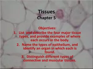
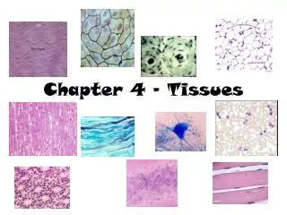
Types of tissues • There are 4 main types of tissues: 1) Epithelial tissue 2) Connective tissue 3) Muscle tissue 4) Nervous tissue
Epithelial Tissue • Functions • Protection • Sensory functions • Secretion • Absorption • Excretion
Epithelial Tissue • Types and locations • Epithelium is divided into two types: • Membranous (covering or lining) epithelium • Glandular epithelium • Locations • Membranous epithelium—covers the body and some of its parts; lines the serous cavities, blood and lymphatic vessels, and respiratory, digestive, and genitourinary tracts • Glandular epithelium—secretory units of endocrine and exocrine glands
Epithelial Tissue • Classification of epithelial tissue • Classification based on cell shape • Squamous • Cuboidal • Columnar • Pseudostratified columnar
Simple epithelium • Simple squamous epithelium • One-cell layer of flat cells • Permeable to many substances • Examples: endothelium—lines blood vessels; mesothelium—pleura
Simple epithelium • Simple cuboidal epithelium • One-cell layer of cuboidal cells • Found in many glands and ducts
Simple epithelium • Simple columnar epithelium • Single layer of tall, column-shaped cells • Cells often modified for specialized functions—e.g., goblet cells (secretion), cilia (movement), microvilli (absorption) • Often lines hollow visceral structures
Simple epithelium • Pseudostratified columnar epithelium • Columnar cells of differing heights • All cells rest on basement membrane but may not reach the free surface above • Cell nuclei at odd and irregular levels • Found lining air passages and segments of male reproductive system • Motile cilia and mucus are important modifications
Stratified epithelium • Stratified squamous (keratinized) epithelium • Multiple layers of flat, squamous cells (Figure 5-9) • Cells filled with keratin • Covers outer skin on body surface
Stratified epithelium • Stratified squamous (nonkeratinized) epithelium (Figure 5-10) • Lines vagina, mouth, and esophagus • Free surface is moist • Primary function is protection
Stratified epithelium • Stratified cuboidal epithelium • Two or more rows of cells are typical • Basement membrane is indistinct • Located in sweat gland ducts and pharynx
Stratified epithelium • Stratified columnar epithelium • Multiple layers of columnar cells • Only most superficial cells are typical in shape • Rare • Located in segments of male urethra and near anus
Stratified epithelium • Stratified transitional epithelium (Figure 5-11) • Located in lining of hollow viscera subjected to stress (e.g., urinary bladder) • Often 10 or more layers thick • Protects organ walls from tearing
Can you identify these? B A E C D F G
Connective Tissue • Functions, characteristics, and types • General function—connects, supports, transports, and protects • General characteristics—extracellular matrix (ECM) predominates in most connective tissues and determines its physical characteristics; consists of fluid, gel, or solid matrix, with or without extracellular fibers (collagenous, reticular, and elastic) and proteoglycans or other compounds that thicken and hold together the tissue
Connective Tissue • Four main types: • Fibrous • Loose, ordinary (areolar) • Adipose • Reticular • Dense • Irregular • Regular (collagenous and elastic) • Bone • Compact bone • Cancellous bone • Cartilage • Hyaline • Fibrocartilage • Elastic • Blood
Fibrous connective tissue • Loose, ordinary (areolar) connective tissue • One of the most widely distributed of all tissues • Intercellular substance is prominent and consists of collagenous and elastic fibers loosely interwoven and embedded in soft, viscous ground substance Function—stretchy, flexible connection
Fibrous connective tissue • Adipose tissue • Similar to loose connective tissue but contains mainly fat cells • Functions—protection, insulation, support, and food reserve
Fibrous connective tissue • Reticular tissue • Forms framework of spleen, lymph nodes, and bone marrow • Consists of network of branching reticular fibers with reticular cells overlying them • Functions—defense against microorganisms and other injurious substances; reticular meshwork filters out injurious particles, and reticular cells phagocytose them
Dense fibrous tissue • Matrix consists mainly of fibers packed densely and relatively few fibroblast cells • Irregular—fibers intertwine irregularly to form a thick mat (Figure 5-20) • Regular—bundles of fibers are arranged in regular, parallel rows • Collagenous—mostly collagenous fibers in ECM (Figure 5-21 and 5-22) • Elastic—mostly elastic fibers in ECM (Figure 5-23) • Locations—composes structures that need great tensile strength, such as tendons and ligaments; also dermis and outer capsule of kidney and spleen • Function—furnishes flexible connections that are strong or stretchy
Bone tissue • Highly specialized connective tissue type • Cells—osteocytes—embedded in a calcified matrix • Inorganic component of matrix accounts for 65% of total bone tissue • Functions: • Support • Protection • Point of attachment for muscles • Reservoir for minerals • Supports blood-forming tissue
Compact bone • Osteon (Haversian system) • Structural unity of bone • Spaces for osteocytes called lacunae • Matrix present in concentric rings called lamellae • Canaliculi are canals that join lacunae with the central Haversian canal • Cell types: • Osteocyte—mature, inactive bone cell • Osteoblast—active, bone-forming cell • Osteoclast—bone-destroying cell • Formation (ossification) (Figure 5-24) • In membranes—e.g., flat bones of skull • From cartilage (endochondral)—e.g., long bones, such as the humerus
Cancellous bone • Trabeculae—thin beams of bone • Supports red bone marrow • Myeloid tissue—a type of reticular tissue • Produces blood cells • Called spongy bone because of its spongelike appearance
Cartilage • Chondrocyte is only cell type present • Lacunae house cells, as in bone • Avascular—therefore, nutrition of cells depends on diffusion of nutrients through matrix • Heals slowly after injury because of slow nutrient transfer to the cells • Perichondrium is membrane that surrounds cartilage
Types of Cartilage • Hyaline (Figure 5-28) • Appearance is shiny and translucent • Most prevalent type of cartilage • Located on the ends of articulating bones • Fibrocartilage (Figure 5-29) • Strongest and most durable type of cartilage • Matrix is semirigid and filled with strong, white fibers • Found in intervertebral disks and pubic symphysis • Serves as shock-absorbing material between bones at the knee (menisci) • Elastic (Figure 5-30) • Contains many fine, elastic fibers • Provides strength and flexibility • Located in external ear and larynx
Blood • A liquid tissue • Contains neither ground substance nor fibers • Composition of whole blood • Liquid fraction (plasma) is the matrix—55% of total blood volume • Formed elements contribute 45% of total blood volume • Red blood cells, erythrocytes • White blood cells, leukocytes • Platelets, thrombocytes
Blood (cont.) • Functions • Transportation • Regulation of body temperature • Regulation of body pH • White blood cells destroy bacteria • Circulating blood tissue is formed in the red bone marrow by a process called hematopoiesis; the blood-forming tissue is sometimes called hematopoietic tissue
Muscle Tissue • Types (Table 5-7) • Skeletal, or striated voluntary (Figure 5-32) • Smooth, or nonstriated involuntary, or visceral (Figures 5-33 and 5-34) • Cardiac, or striated involuntary (Figure 5-35)
Muscle Tissue • Microscopic characteristics • Skeletal muscle—threadlike cells with many cross striations and many nuclei per cell • Smooth muscle—elongated, narrow cells, no cross striations, one nucleus per cell • Cardiac muscle—branching cells with intercalated disks (formed by abutment of plasma membranes of two cells)
Nervous Tissue • Functions—rapid regulation and integration of body activities • Specialized characteristics • Excitability • Conductivity • Organs • Brain • Spinal cord • Nerves
Nervous Tissue • Cell types (Table 5-7) • Neuron—conducting unit of system (Figure 5-36) • Cell body, or soma • Processes • Axon (single process)—transmits nerve impulse away from the cell body • Dendrites (one or more)—transmit nerve impulse toward the cell body and axon • Neuroglia—special connecting, supporting, coordinating cells that surround the neurons
Tissue Repair • Tissues have a varying capacity to repair themselves; damaged tissue regenerates or is replaced by scar tissue • Regeneration—growth of new tissue (Figure 5-37) • Scar—dense fibrous mass; unusually thick scar is a keloid (Figure 5-38) • Epithelial and connective tissues have the greatest ability to regenerate • Muscle and nervous tissues have a limited capacity to regenerate