Exploring Human Tissues: Epithelial and Connective Tissue Types
Dive into the world of tissues with a focus on epithelial and connective types, their structures, functions, and diverse locations in the human body. Discover the composition, fibers, and ground substance that make up connective tissues.

Exploring Human Tissues: Epithelial and Connective Tissue Types
E N D
Presentation Transcript
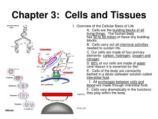
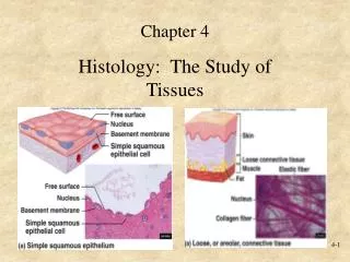
Histology The study of tissues =
There are four major tissue types in the body: • Epithelial • Muscle • Nerve • Connective
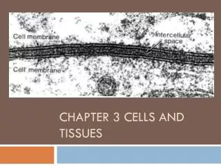
A) Locations • covers all body surfaces – inside and outside • forms inner lining of body cavities and organs
B) Structure • Damaged cells are continually replaced. • Cells are tightly packed and often attached to one another by desmosomes & tight junctions • Substances readily pass between cells • e.g. gap junctions
C) Functions of Epithelial Tissue • Protection • Absorption • Filtration • Secretion
1. Simple Squamous Epithelial • Absorption • diffusion • filtration
Absorption • Secretion • liver • thyroid • kidney tubules Cross-Section of a kidney tubule
3. Simple Columnar Epithelial • Absorption • Movement (e.g. egg and embryo move along uterine tube)—by cilia at cell surface • Secretion (e.g. goblet cells in the large intestine secrete mucus to ease the passage of feces.)
4. Stratified Squamous Epithelium • multi-layered protection
protects against mechanical abrasion, water loss, and pathogen entry e.g. sole of the foot
5. Stratified Cuboidal Epithelium • Protection e.g. sweat gland ducts, mammary glands
6. Pseudostratified Columnar Epithelium • looks multi-layered, but it’s NOT! • cells are of varying heights , and nuclei are at several levels. • goblet cells present • often ciliated at the surface
Here, we have pathogens traveling down the pharynx trying to attack the surface cells. How can they be repelled???
Ahhh!!! It’s so sticky and slimy! The mucosal cells lining the trachea release a flood of mucus, trapping the pathogens!
blub, blub . . . . The cilia successfully sweep the pathogens up and away!
7. Transitional Epithelium • resembles stratified squamous epithelium, but surface cells are rounded and often bulge above surface (dome-shaped)
distensible e.g. urinary bladder and tract lining
Connective Tissue • found throughout the body, and as parts of various organs • composed of connective tissue cells + extracellular matrix • extracellular matrix = fibers + ground substance [fluid-solid]
Functions: • protects internal organs, provide support, connect organs together • provide framework • fill spaces • produce blood cells • provide immune protection • tissue repair
Composition 1. Fibers Collagen Fibers as seen with a scanning electron microscope
a) collagenous fibers • interwoven strands of collagen (protein) • most abundant protein • thick fibers with great tensile strength • white & wavy appearance e.g. tendons, ligaments, deep layer of the skin Close-up of a single fiber
b) elastic fibers • made of elastin protein • coiled structure to stretch and snap back like a rubber band • e.g. lungs, arteries, and skin
c) reticular fibers • very thin collagen fibers that branch extensively • form frameworks for organs e.g. liver
2. Ground substance • Gelatinous material between the connective tissue cells and fibers [fluid to semi-solid to solid]
a) areolar connective tissue 1) Loose Connective Tissue • Loose arrangement of all 3 fiber types
: e.g. under epithelia, surrounding blood vessels, nerves, between muscles
b) reticular tissue • loose network of reticular fibers • form a scaffold-like framework for lymphatic organs. e.g. lymph nodes, spleen
c) adipose tissue • adipocytes – large, clear cells with thin margins. • Subcutaneous fat beneath skin, breast, heart, eyes
Functions of adipose tissue: • Energy storage • Thermal insulation • Shock absorption • Protective cushioning
a) Dense Regular CT 2. Dense Connective Tissue • Densely packed, parallel, often wavy collagenous fibers • Little open space e.g. ligaments bind bone to other bones tendons attach skeletal muscles to bone