Chapter 5 Tissues
Chapter 5 Tissues. Intercellular Connections . Individual cells connect to form tissues 3 ways: Tight junctions- prevents permeability of ions through junction. Desmosome- adhesion between cells in spots. Allows from some permeability.


Chapter 5 Tissues
E N D
Presentation Transcript
Intercellular Connections • Individual cells connect to form tissues 3 ways: • Tight junctions- prevents permeability of ions through junction. • Desmosome- adhesion between cells in spots. Allows from some permeability. • Gap junctions- cytoplasms of adjacent cells are connected through transport proteins. • Ions can pass freely through cells.
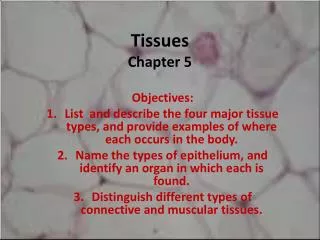
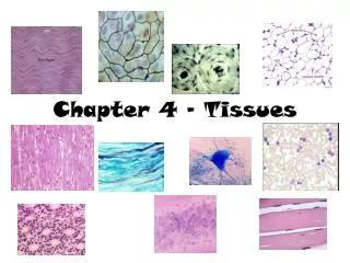
Tissue Types A tissue is a group of cells with a common structure & function The human body is composed of four main tissue types: • 1. Epithelial • 2. Connective • 3. Muscle • 4. Nerve
Epithelial Tissue Epithelial Tissue
Characteristics • Always has a free (apical) surface exposed to outside or open space. • Lacks blood vessels • Readily divide and replicate • Has a basement membrane to anchor underlying tissue • Between epithelial and connective tissue
Functions • Covers body surfaces • Makes up glands • Protects • Absorbs • Secretes • Excretes
Classified by Shape • Squamous – flattened cells • Cuboidal – cube-shaped • Columnar – tall, column-shaped
Classified by Shape May occur in layers: • Simple – 1 layer of cells • Stratified – 2 or more layers • Pseudostratified – appears to be layered, but is not • Example – simple cuboidal • Example – stratified columnar
s Simple Squamous- Thin, flattened cells. Allow for diffusion and filtration. Line air sacs of lungs and walls of capillaries.
Simple cuboidal-single layer of cube shaped cells. Lines follicles of thyroid gland, kidneys and ducts of certain glands. Used for secretion and absorption
Simple columnar- single layer of elongated cells. Can contain cilia, used for protection and absorption in digestive tract. Can contain goblet cells.
Stratified squamous-Layers of squamous cells. Make up epidermis and line cavities exposed to external environment. Outer layer die and accumulate keratin.
Stratified columnar- Several layers of columnar cells overlying cuboidal cells near the basement membrane. Found in male reprod. System and pharynx
Pseudostratified ciliated columnar- Appear stratified but are not. Often contain cilia and goblet cells which secrete mucus. Line respiratory passages.
Pseudostratified ciliated columnarw/goblet cells- Line Respiratory passages to trap unwanted particles
Transitional tissue- Changes in response to change in tension (stretching). Line urinary bladder and urethra. Larger cells at surface, smaller cells deeper.
Glandular Epithelium • Specialized to secrete substances • Usually glands are lined w/cuboidal or columnar epithelium • Those that secrete substances into ducts that open onto a surface are exocrine glands (salivary, oil glands, etc.) • Those that secrete into tissues or bloodare endocrine glands (pituitary)
Classifying Glands by Structure • Simple- does not branch off before reaching secretory portion • Compound- duct that does branch before secretory portion.
Classifying Glands by Type of Secretions 3 types: • Small portions of cells • in secretions • No loss of cytoplasm Ex. – mammary glands in secretions • Ex. – pancreas
Classifying by Secretions • Secretions w/entire cells filled w/secretory products; ex. – sebaceous (oil) glands
Functions • 1. connects • 2. supports • 3. protects • 4. provides framework • 5. fills spaces
Functions • 6. stores fat • 7. produces blood cells • 8. protects against infection • 9. transports nutrients • 10.helps repair damaged tissue
Characteristics • 1. Consists of cells in a matrix (intercellular material) • 2. Cells some distance apart • 3. Most can divide • 4. Good blood supply
Types of Fibers: • collagenous – composed of collagen (protein); have great tensile strength; slightly elastic; compose bones, tendons & ligaments
Types of Fibers - continued • elastic – composed of elastin (protein); very elastic but weaker; compose vocal cords & air passages of lungs
Types of Fibers - continued • Reticular – composed of very fine collagenous fibers. • Form support network.
Types of Cells 1. Fixed cells – stay in one place & have stable numbers; 2 types: • fibroblasts – large & star-shaped; most prevalent
Types of Cells - continued • mast cells – may release heparin (for blood clotting) & histamines (promotes allergic reactions & inflammation); usually located near blood vessel walls
Types of Cells - continued 2. Wandering cells – • macrophages – travel through body; numbers change in response to infection; scavengers (Purple cells – macrophages, Green cells – T-lymphocytes)
Areolar tissue-binds the skin to underlying organs and under epithelium to provide bloodflow. Binds and protects. A- fibroblast, B-collagen, C- elastin
Adipose tissue- connective tissue composed of fats, cushion joints and provide insulation. A- nuclei, B- fat globules
Regular dense connective- strong fibers bind body parts together. Found in ligaments and tendons. Poor blood supply so slow healing. A- fibroblasts. B- collagen and elastin
Irregular dense connective- disorganized and strong. Found in the dermis
Hyaline cartilage- Most common, found on ends of bones, nose cavity and supporting rings of resp. system. A- chondrocytes, B- Matrix (fine collagen fibers), C- Lacunae
Fibrocartilage- tough tissue containing collagenous fibers. Shock absorbers between vertebrae and pubic girdle. A- Chondrocyte, B- Collagen fibers
Elastic cartilage- flexible cartilage make up ears and larynx. Used for flexible support. A- chondrocytes, B- elastin, C- Lacunae
Blood – red cells & white cellRed- used for transport, white- immunity
Elastic connective- allows for stretching, found in attachments between vertebrae. A- elastic fibers
Reticular connective- walls of liver and spleen. Used for support.
Bone- A- central canal (contains blood vessels) B- Canaliculi- minute tubes allow for movement between cells.
Bone- D- Lamellae (layers of osetocytes), C- osteocytes (Bone Cells)
Muscle Tissue 3 types: • Skeletal- Attached to bone and controlled by conscious effort (Voluntary). • Used for movement • Striated • Long and thin with multiple nuclei
Muscle Tissue Cont. • Smooth- lacks striations found in skeletal, used for involuntary movements • Ex- move food through digestive tract • Cardiac- striated muscle found only in the heart • At intercellular junction contain intercalated discs. • Allows for heart to contract as one unit