Tissues: Types and Functions
Explore the principal types of tissues including epithelial, connective, muscle, and nervous tissues, along with their extracellular matrix components and functions. Learn about the classification, functions, and examples of epithelial tissue.


Tissues: Types and Functions
E N D
Presentation Transcript
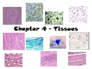
Principal Types of Tissue (Table 5-1) • Epithelial tissue – covering and lining • Connective tissue – specialized to support the body and its parts, connect and hold them together, transport substances through the body, and protect it from foreign invaders • Muscle tissue – produces movement, has cells that are specialized to contract • Nervous tissue – conductive tissue that communicates between various parts of the body
Extracellular Matrix (ECM) • Complex, nonliving material between cells in a tissue (Figure 5-1) • Some tissues have a large amount of ECM; other tissues have hardly any ECM • Different kinds of components give ECM in different tissues a variety of characteristics
Extracellular Matrix (ECM) • Components (Table 5-2) • Water • Proteins • Structural proteins • Collagen—strong, flexible protein fiber • Elastin—elastic fibers • Glycoproteins (Fibronectin & Laminin) - proteins with a few carbohydrate attachments • Proteoglycans • Hybrid molecules that are mostly carbohydrates attached to a protein backbone
Extracellular Matrix (ECM) • Functions • Helps bind tissues together structurally • ECM components bind to each other and to integrins in plasma membranes of cells • In some tissues, it is primarily intercellular junctions that hold cells together • Allows local communication among ECM and various cells—through connection via integrins in plasma membranes
Epithelial Tissue • Types and locations • Epithelium is divided into two types: • Membranous (covering or lining) epithelium • Glandular epithelium • Locations • Membranous epithelium—covers the body and some of its parts; lines the serous cavities, blood and lymphatic vessels, and respiratory, digestive, and genitourinary tracts • Glandular epithelium—secretory units of endocrine and exocrine glands
Epithelial Tissue • Functions • Protection – tough and impermeable, protects the body from mechanical and chemical injury and invading bacteria and other disease causing microorganisms • Sensory functions – structures that are specialized for sensation found around the skin, nose, eye and ear • Secretion – secretion products include: hormones, mucus, digestive juices, and sweat • Absorption – lining of the gut and exchange of gases in the lungs • Excretion – tubules in the kidneys
Epithelial Tissue • Generalizations about epithelial tissue • Limited intercellular matrix • Basement membrane attaches epithelial tissue to an underlying connective tissue layer • Avascular • Cells are in close proximity, with many desmosomes and tight junctions • Capable of reproducing itself, highly mitotic
Epithelial Tissue • Membranous (covering or lining) epithelium (Table 5-3) • Classification based on cell shape • Squamous • Cuboidal • Columnar • Pseudostratified columnar
Epithelial Tissue • Classification of epithelial tissue based on layers • Classifications based on layers of cells • Simple • Stratified • Transitional
Epithelial Tissue • Simple epithelium • Simple squamous epithelium (Figures 5-4 and 5-5) • One-cell layer of flat cells • Permeable to many substances • Examples: O2 & CO2 exchange in the lungs, movement of fluid and dissolved substances between the blood and across blood vessels by osmosis and filtration in the kidneys • Simple cuboidal epithelium (Figure 5-6) • One-cell layer of cuboidal cells • Found in many glands and ducts • Examples: secretion of substances such as tears and saliva, and in absorption, such as re-absorption of water by kidney cells
Epithelial Tissue • Simple epithelium (cont.) • Simple columnar epithelium (Figure 5-7) • Single layer of tall, column-shaped cells • Often lines hollow visceral structures • Nonciliated simple columnar epithelium – microvilli and goblet cells. Microvilli – finger like projections that increase surface area. Goblet Cells – secrete mucous. Line the G.I., Respiratory, Reproductive, and Urinary Tracts. • Ciliated simple columnar epithelium – cilia, line the upper respiratory tract, move particles towards the throat. Line the fallopian tubes. • Pseudostratified columnar epithelium (Figure 5-8) • Columnar cells of differing heights • Give the impression of a multi-layered tissue, this is why they are given the name • Found lining air passages of upper respiratory and segments of male reproductive system • Can have cilia or can have none • There function is secretion and movement of mucous by ciliary action
Epithelial Tissue • Stratified epithelium • Stratified squamous (keratinized) epithelium • Multiple layers of flat, squamous cells (Figure 5-9) • Cells filled with keratin • Covers outer skin on body surface • Stratified squamous (nonkeratinized) epithelium (Figure 5-10) • Lines vagina, cervix, mouth, and esophagus • Free surface is moist • Primary function is protection • Pap smear
Epithelial Tissue • Stratified epithelium (cont.) • Stratified cuboidal epithelium • Two or more rows of cells are typical • Located in sweat gland ducts and pharynx • Stratified columnar epithelium • Multiple layers of columnar cells • Only most superficial cells are typical in shape • Rare • Located in segments of male urethra and near anus
Epithelial Tissue • Stratified epithelium (cont.) • Stratified transitional epithelium (Figure 5-11) • Located in lining of hollow viscera subjected to stress (e.g., urinary bladder) • Often 10 or more layers thick • Protects organ walls from tearing
Epithelial Tissue • Glandular epithelium • Specialized for secretory activity • Exocrine glands—discharge secretions into ducts that empty at the surface of covering and lining epithelium or directly onto a free surface. • secretions include mucus, perspiration, oil, wax, and digestive enzymes, and salivary glands. • Endocrine glands—“ductless” glands; discharge secretions directly into the blood or interstitial fluid. The thyroid and pituitary glands are examples.
Connective Tissue • Functions • Connect – connects tissues and muscles to each other, muscles to bones, and bones to bones • Supports – forms a supporting framework for the body and its organs • Transports – bloods components to cells in the body • Defends – fend off foreign invaders and other microrganisms
Connective Tissue • Cells of CT • Fibroblasts - secrete a gel-like substance that forms the ground substance and fibers of the matrix. They are present in all connective tissue. • Macrophages - engulf bacteria by phagocytosis • Plasma Cells - secrete antibodies, which are proteins that attack or neutralize foreign substances in the body. • Mast Cells - secrete heparin, prostaglandins, and histamine (chemical that dilates small blood vessels during inflammation or injury). • Adipocytes – store fat • Leukocytes (white blood cells) – not found in significant numbers in tissue, but can migrate from the blood to the tissue.
Connective Tissue • Four main types (Table 5-6): • Fibrous • Loose, ordinary (areolar) • Adipose • Reticular • Dense • Irregular • Regular (collagenous and elastic) • Bone • Compact bone • Cancellous bone • Cartilage • Hyaline • Fibrocartilage • Elastic • Blood
Connective Tissue • Fibrous connective tissue • Loose, ordinary (areolar) connective tissue (Figure 5-15) • One of the most widely distributed of all tissues • Intercellular substance is prominent and consists of collagenous and elastic fibers loosely interwoven and embedded in a soft, viscous ground substance • Several kinds of cells present: notably, fibroblasts and macrophages; also mast cells, plasma cells, fat cells, and some white blood cells (Figure 5-16) • Function—stretchy, flexible connection
Connective Tissue • Adipose tissue (Figures 5-17 and 5-18) • Similar to loose connective tissue but contains mainly fat cells (adipocytes) • Functions—protection, insulation, support, and food reserve • Reticular tissue (Figure 5-19) • Forms framework of spleen, lymph nodes, and bone marrow • Consists of network of branching reticular fibers with reticular cells overlying them • Functions—defense against microorganisms and other injurious substances; reticular meshwork filters out injurious particles, and reticular cells phagocytose them
Inflammation • Inflammatory Response – p. 167
Connective Tissue • Dense fibrous tissue • Matrix consists mainly of fibers packed densely and relatively few fibroblast cells • Irregular—fibers intertwine irregularly to form a thick mat, found in areas of the body where tensions are exerted in various directions • Dermis, heart valves, periosteum • Regular—bundles of fibers are arranged in regular, parallel rows • Tendons and ligaments • Elastic—mostly elastic fibers in ECM (Figure 5-23), provides stretch and strength • Lungs and arteries • Locations—composes structures that need great tensile strength, such as tendons and ligaments; also dermis and outer capsule of kidney and spleen • Function—furnishes flexible connections that are strong or stretchy
Connective Tissue • Bone tissue • Highly specialized connective tissue type • Cells—osteocytes—embedded in a calcified matrix • Inorganic component of matrix accounts for 65% of total bone tissue • Functions: • Support • Protection • Point of attachment for muscles • Reservoir for minerals • Supports blood-forming tissue
Connective Tissue • Compact bone (Figures 5-25 and 5-26) • Osteon (Haversian system) • Structural unit of bone • Spaces for osteocytes called lacunae • Matrix present in concentric rings called lamellae • Canaliculi are canals that join lacunae with the central Haversian canal • Cell types: • Osteocyte—mature, inactive bone cell • Osteoblast—active, bone-forming cell • Osteoclast—bone-destroying cell • Formation (ossification) (Figure 5-24) • In membranes—e.g., flat bones of skull • From cartilage (endochondral)—e.g., long bones, such as the humerus
Connective Tissue • Cancellous bone (Figures 5- 25 and 5-27) • Trabeculae—thin beams of bone • Supports red bone marrow • Myeloid tissue—a type of reticular tissue • Produces blood cells • Called spongy bone because of its spongelike appearance
Connective Tissue • Cartilage • Chondrocyte is the only cell type present • Lacunae house cells, as in bone • Avascular—therefore, nutrition of cells depends on diffusion of nutrients through matrix • Heals slowly after injury because of slow nutrient transfer to the cells
Connective Tissue • Types • Hyaline (Figure 5-28) • Appearance is shiny and translucent • Most prevalent type of cartilage • Located on the ends of articulating bones • Fibrocartilage (Figure 5-29) • Strongest and most durable type of cartilage • Matrix is semirigid and filled with strong, white fibers • Found in intervertebral disks and pubic symphysis • Serves as shock-absorbing material between bones at the knee (menisci) • Elastic (Figure 5-30) • Contains many fine, elastic fibers • Provides strength and flexibility • Located in external ear and larynx
Connective Tissue • Blood • A liquid tissue (Figure 5-31) • Contains neither ground substance nor fibers • Composition of whole blood • Liquid fraction (plasma) is the matrix—55% of total blood volume • Formed elements contribute 45% of total blood volume • Red blood cells, erythrocytes • White blood cells, leukocytes • Platelets, thrombocytes
Connective Tissue • Blood (cont.) • Functions • Transportation • Regulation of body temperature • Regulation of body pH • White blood cells destroy bacteria • Circulating blood tissue is formed in the red bone marrow by a process called hematopoiesis; the blood-forming tissue is sometimes called hematopoietic tissue
Muscle Tissue • Types (Table 5-7) • Skeletal, or striated voluntary (Figure 5-32) • Smooth, or nonstriated involuntary, or visceral (Figures 5-33 and 5-34) • Cardiac, or striated involuntary (Figure 5-35) • Microscopic characteristics • Skeletal muscle—threadlike cells with many cross striations and many nuclei per cell • Smooth muscle—elongated, narrow cells, no cross striations, one nucleus per cell • Cardiac muscle—branching cells with intercalated disks (formed by abutment of plasma membranes of two cells)
Nervous Tissue • Functions—rapid regulation and integration of body activities • Specialized characteristics • Excitability • Conductivity • Organs • Brain • Spinal cord • Nerves
Nervous Tissue • Cell types (Table 5-7) • Neuron—conducting unit of system (Figure 5-36) • Cell body, or soma • Processes • Axon (single process)—transmits nerve impulse away from the cell body • Dendrites (one or more)—transmit nerve impulse toward the cell body and axon • Neuroglia—special connecting, supporting, coordinating cells that surround the neurons
Tissue Repair • Tissues have a varying capacity to repair themselves; damaged tissue regenerates or is replaced by scar tissue • Regeneration—growth of new tissue (Figure 5-37) • Scar—dense fibrous mass; unusually thick scar is a keloid (Figure 5-38) • Epithelial and connective tissues have the greatest ability to regenerate • Muscle and nervous tissues have a limited capacity to regenerate
Body Membranes • Membranes are thin tissue layers that cover surfaces, line cavities, and divide spaces or organs (Figure 5-39, Table 5-8) • Epithelial membranes are the most common type (Figure 5-40) • Cutaneous membrane (skin) • Primary organ of integumentary system • One of the most important organs • Composes approximately 16% of body weight • Serous membrane (serosa) • Parietal membranes—line closed body cavities • Visceral membranes—cover visceral organs • Pleura—surrounds a lung and lines the thoracic cavity • Peritoneum—covers the abdominal viscera and lines the abdominal cavity