Download
1 / 17
170 likes | 211 Vues
Learn about antigen presentation, processing pathways, MHC molecules, and types of antigen-presenting cells such as dendritic cells, macrophages, and B cells. Discover how antigens are recognized and presented to T cells.

E N D
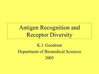
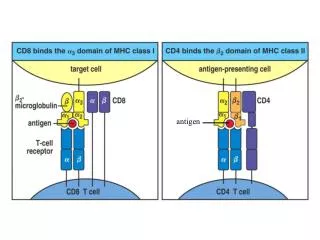
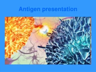
The schematic representation of the interaction of the class II MHC, processed peptide antigen, and T cell receptor molecules during antigen presentation.
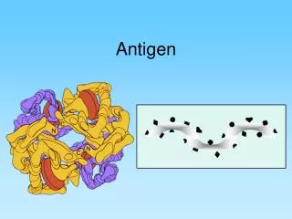
Epitope : That part of an antigen recognized by an antigen receptor (antigenic determinant). Paratope :The part of T cell receptor or antibody that recognizes the part of an antigen. Desetope: The region of class II MHC molecules that reacts with the antigen during antigen presentation. Agretope: The region of a protein antigen that combines with an MHC class II molecule during antigen presentation. Histotope: The portion of an MHC class II molecule that reacts with a T lymphocyte receptor. Restitope: The segment of a T cell receptor that makes contact and interacts with a class II molecule during antigen presentation.
Antigen Processing and Presentation • Antigen processing: refers to the ability of APCs to break down a protein antigen into peptides and to associate those peptides with MHC molecules. • Antigen presentation: is the process of displaying peptide antigens associated with MHC molecules to a T cell. • The path leading to the association of protein fragments with MHC molecules differs for class I and class II MHC. • MHC class I molecules present degradation products derived from intracellular (endogenous) proteins in the cytosol. • MHC class II molecules present fragments derived from extracellular (exogenous) proteins that are located in an intracellular compartment.
Antigen Processing and Presentation Class I MHC Pathway • All nucleated cells express class I MHC. • Proteins are fragmented in the cytosol by proteosomes (a complex of proteins having proteolytic activity) or by other proteases. • The fragments are then transported across the membrane of the endoplasmic reticulum by transporter proteins. (The transporter proteins and some components of the proteosome are encoded by genes in the MHC complex). s s s s s s s s
Antigen Processing and Presentation Class I MHC Pathway (contd.) • Synthesis and assembly of class I heavy chain and β2- microglobulin occurs in the endoplasmic reticulum. • Within the endoplasmic reticulum, the MHC class I heavy chain, β2- microglobulin and peptide form a stable complex that is transported to the cell surface.
Antigen Processing and Presentation Class II MHC Pathway • Only a limited group of cells express class II MHC, which includes the antigen presenting cells (APC). • The principal APC are macrophages, dendritic cells (Langerhans cells), and B cells. • The expression of class II MHC molecules is either constitutive or inducible, especially by interferon- gamma in the case of macrophages.
Antigen Processing and Presentation Class II MHC Pathway (cont.) • Exogenous proteins taken in by endocytosis are fragmented by proteases in an endosome. • The alpha and beta chains of MHC class II, along with an invariant chain, are synthesized and assembled in the endoplasmic reticulum. • The invariant chain prevents endogenous peptides from the cytosol from associating with class II MHC molecules.
Antigen Processing and Presentation Class II MHC Pathway (cont.) • The class II MHC molecules with the associated invariant chain are transported through the Golgi body to reach the endosome. • The invariant chain is digested after reaching to the endosome. • The peptide fragments from the exogenous protein are able to associate with the class II MHC molecules, which are finally transported to the cell surface.
Antigen presenting cells (APC) • Cells with the capacity to capture, process and present antigenic peptides to T cells • Antigens are presented in the context of MHC class I or II • Also deliver co-stimulatory signal (signal II) to T cells leading to proper activation • Only APCs can activate a naïve T cell The three main types of antigen presenting cells are: Dendritic cells, Macrophages, B cells
Antigen presenting cells (APC) Dendritic cells: • found in skin and other tissues, ingest antigens by pinocytosis and transport antigens to the lymph nodes and spleen. • In the lymph nodes and spleen they are found predominantly in the T cells areas. • Dendritic cells are the most effective antigen presenting cells and can present antigens to naïve (virgin) T cells. • Furthermore, they can present internalized antigens in association with either class I or class II MHC molecules (cross presentation), although the predominant pathway for internalized antigen is the class II pathway.
Antigen presenting cells (APC) Macrophages: • The second type of antigen presenting cell. • These cells ingest antigen by phagocytosis or pinocytosis. Macrophages are not as effective in presenting antigen to naïve T cells but they are very good in activating memory T cells. B cell: • The third type of antigen presenting cell is the B cell. • These cells bind antigen via their surface Ig and ingest antigens by pinocytosis. • Like macrophages these cells are not as effective as dendrite cells in presenting antigen to naïve T cells. • B cells are very effective in presenting antigen to memory T cells, especially when the antigen concentration is low because surface Ig on the B cells binds antigen with a high affinity.
Cell-Cell Interactions in Specific Immune Responses • To discuss the central role of Th cells in immune responses. • To describe the cell-cell interactions which occur in • (i) antibody responses to T-dependent antigens, • (ii) generation of cytotoxic T cells, and • (iii) activation of macrophages and NK cells. • 3. To discuss the mechanisms of killing by cytotoxic T cells and NK cells • 4. To discuss responses to T-independent antigens.
I. Central role of Th cells in immune responses • After Th cells recognize specific • antigen presented by an APC, they can initiate several key immune processes. • These include: • 1) selection of appropriate • effector mechanisms ( e.g., B • cell activation or T generation) • 2) induction of proliferation of • appropriate effector cells and • 3) enhancement of the functional • activities of other cells (e.g., • granulocytes, macrophages, NK • cells). Central role of Th cells in immune responses.
I. Central role of Th cells in immune responses • There are three subpopulations of Th cells, Th0, Th1 and Th2 cells. • When naïve Th0 cells encounters antigen in secondary lymphoid tissues, they are capable of differentiating into inflammatory Th1 cells or a helper Th2 cells, which are distinguished by the cytokines they produce. • Whether a Th0 cells becomes a Th1 or aTh2 cell depends upon the cytokines in the environment, which is influenced by antigen. For example some antigens stimulate IL-4 production which favors the generation of Th2 cells while other antigens stimulate IL-12 production, which favors the generation of Th1 cells. Differentiation of Th cells.
I. Central role of Th cells in immune responses Th1 and Th2 cells affect different cells and influence the type of an immune response. • Cytokines produced by Th1 cellsactivate macrophages and participate in the generation of Tc cells, resulting in a cell-mediated immune response. • Cytokines produced by Th2 cells help to activate B cells, resulting in antibody production. • Th2 cytokines also activate granulocytes. • Each subpopulation can also exert inhibitory influences on the other: IFN-γ produced by Th1 cells inhibits proliferation of Th2 cells and Il-10 produced by Th2 cells inhibits production of IFN-γ by Th1 cells. Selection of effector mechanisms by Th1 and Th2 cells.