Download
1 / 43
430 likes | 811 Vues
This presentation will discuss single-crystal diffraction in biology, covering topics such as resolution, crystallography, experiment formalism, lattice symmetries, and intensity relationships. Learn about crystal growth, Bragg’s law, Laue conditions, and 3D space vectors in the context of determining 3D structures.

E N D
This presentation will probably involve audience discussion, which will create action items. Use PowerPoint to keep track of these action items during your presentation • In Slide Show, click on the right mouse button • Select “Meeting Minder” • Select the “Action Items” tab • Type in action items as they come up • Click OK to dismiss this box • This will automatically create an Action Item slide at the end of your presentation with your points entered. Introduction toSingle-crystal Diffraction Biology 555 Andrew J. Howard 11 September 2018
Diffraction: why Resolution Crystallography: how Experiment Formalism Lattice Symmetries Cell matrices Intensity relationships Images and measurements Rotation vs. Laue What we’ll discuss Single-crystal diffraction I
Determining 3-D structures • We want to know the structures of macromolecules because we can understand function on the basis of structure • If we already know what the function is, we use the structure to figure out how it works • If we don’t know the function, we sometimes can use the structure to figure out what the function is. Single-crystal diffraction I
How do we do this? • If we already know something about structure, we can use a variety of techniques (e.g. light scattering, X-ray absorption, EPR , fiber diffraction) to answer specific structural questions • If we don’t know anything: • Low resolution (details > 3Å):cryoEM, CD, SAXS, SANS • High resolution (details < 3Å) • single-crystal diffraction • multidimensional NMR Single-crystal diffraction I
What does resolution mean? • In general it refers to the distance associated with the smallest detail discernible from a particular experiment or approach • Specific significance depends on the technique involved • In crystallography it’s related to the widest angle of diffraction measured, via Bragg’s law Single-crystal diffraction I
Crystallography Determine 3-D structure by: • Making a well-ordered 3-D crystal • Identifying Bragg diffraction spots • Measuring their intensities • Correcting for systematic errors • Getting phase angles experimentally or by indirect reasoning • Calculating electron density by Fourier techniques • Relating electron density to atomic positions Single-crystal diffraction I
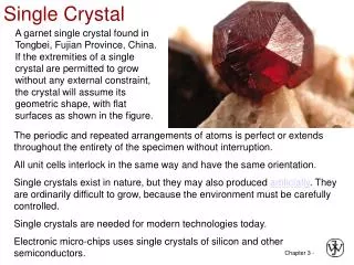
Crystal growth • We’ll discuss this in detailTuesday • Recognize at the outset that protein crystals are artificial constructs: very few proteins naturally form 3-D ordered arrays • But there are systematic ways to induce crystallization Single-crystal diffraction I
What does 3-D order do? • Briefly, we say that the 3-D order collapses what would otherwise be a fuzzy scattering pattern into a series of discrete spots • Each spot has 3 integer indices that characterize it (Why? 3 dimensions) • If we can identify each spot (determine the 3 integers) and measure the intensity of it, we have the Fourier amplitudes needed in the transform. Single-crystal diffraction I
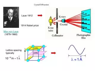
How does the experiment work? • Last time we showed a simple-minded derivation of Bragg’s law based on planes of diffracting objects • This is an oversimplificationbut it yields nl = 2Dsinq • n = integer order of plane • l = incoming X-ray wavelength • D = spacing between planes • q = angle between diffracting plane and incoming or outgoing beam direction Single-crystal diffraction I
Bragg’s law cartoon Image courtesy Mineral Physics Institute, SUNY Stonybrook Single-crystal diffraction I
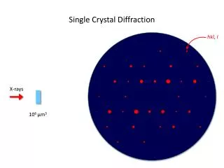
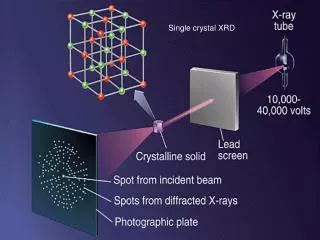
Typical diffraction image • Obtained with monochromatic X-rays and with the crystal approximately stationary • This doesn’ttell the whole story of crystal diffraction, but it’s a start. Single-crystal diffraction I
How would a small-molecule crystallographer react to that? • Too many spots! • In a typical small-molecule experiment with the crystal stationary, the Laue conditions are only satisfied for a very few Bragg spots—maybe 3. • So the small-molecule person would be impressed that hundreds of spots are visible in a still image Single-crystal diffraction I
Laue conditions for Bragg diffraction spots Laue condition for 3-D crystals tells us: • Bragg diffraction occurs in discrete angular directions (rays spots) • Each spot can be identified with three integer indices (h,k,l), e.g. (-6,11,-4) • For a given sample orientation, Laue condition is only satisfied for a few index values Single-crystal diffraction I
3-space vectors • Get used to thinking about ordered triples of numbers as vectors • That’s natural with things like diffraction vectors, since they exist in a three-space • But we can even treat a triple of reflection indices (h,k,l) as a vector h. • Convention: boldface lowercase letters (e.g. s, k, h) denote vectors; boldface capital letters (e.g. A, R) denote square matrices Single-crystal diffraction I
What will we use this for? • We’re shooting electromagnetic waves exp(2pik0•x) at a sample • Scatter with amplitude exp(2pik•x) • Difference=exp(2pis•x) for s = k - k0 • I ~ |F|2 = [Sx f(x) exp(i(k•x-k0•x))]2 -k0 k s = k-k0 k k0 Single-crystal diffraction I
Using these structure factors to determine a structure • We need all of these structure factors to determine electron densities: • r(r) = ShklFhkl exp(-2pi(s•r)) • Thus the electron density is the inverse Fourier transform of the structure factors • This is a triple sum over the three integer indices h = (h,k,l) • In principle it extends from -∞ to ∞ in all three indices; in practice it covers a narrower range Single-crystal diffraction I
Relating s to h • The diffraction vector s is related to the index vector h in a simple way: • s = lA•hwhere A is a 3x3 matrix describing the reciprocal-space unit cell associated with the real sample • This is a concept we’ll revisit later Single-crystal diffraction I
Relating h to s • A more intuitive description: • h = R•s / lwhere the index vector h(e.g. (6,-11,4)) is related to the diffraction vector s via a 3x3 matrix R describing the real-space unit cell • Columns of R = unit cell lengths a, b, c • Dot products between columns yield the unit cell angles a, b, g • Note that R = A-1 Single-crystal diffraction I
Sample symmetry and R • The rotational symmetry of the unit cell gives rise to specific properties of the unit cell matrix R • If a=g=90º,b ≠90º, it’s monoclinic • a=b=g=90º, lengths unrestricted:orthorhombic • a=b=g=90º, a=b: tetragonal • a=b=90º, g=120º, a=b: hexagonal or trigonal • a=b=c, a=b=g: rhombohedral • a=b=c, a=b=gº: cubic • Those are the only possibilities, apart from redefinitions of what we mean by a, b, c. Single-crystal diffraction I
Crystal systems I,II www.dkimages.com • Triclinica ≠ b ≠ c ≠ ≠ ≠90º • Monoclinica ≠ b ≠ c = = 90º ≠ 90º Single-crystal diffraction I
Crystal Systems III, IV • Orthorhombica ≠ b ≠ c = = = 90º • Tetragonala = b ≠c = = = 90º Single-crystal diffraction I
Crystal systems V, VI • Cubica = b = c = = = 90º • Hexagonala = b ≠c = = 90º = 120º Single-crystal diffraction I
Crystal System 7 • Rhombohedrala = b = c = = ≠ 90º Single-crystal diffraction I
Crystal systems and symmetries • Crystals must have 3-D translational order or they aren’t crystals • They may have rotational symmetries within the unit cell • The crystal systems are specifically associated with those symmetries Single-crystal diffraction I
Kinds of symmetries • Rotations:2-fold, 3-fold,4-fold, 6-fold • Screw axes: rotation combined with a fractional translation • 2-fold: half a unit cell • 3-fold: 1/3 or 2/3 (=-1/3) • 4-fold: 1/4, 1/2, or 3/4 (=-1/4) • 6-fold: 1/6, 1/3, 1/2, 2/3 (-1/3), 5/6 (=-1/3) Single-crystal diffraction I
Other symmetries • Centers of symmetry: r -r • Mirrors: you can figure that out; A mirror through Y maps(x, y, z) into (x, -y, z) • Glide planes: center of symmetry combined with translation Single-crystal diffraction I
Specific symmetries • Triclinic: none • Monoclinic: twofold axis* about b • Orthorhombic: twofold axis*about a, b, c • Tetragonal: fourfold axis* about c • Cubic: threefold axis down body diagonal; twofold axis* perpendicular • Hexagonal: threefold* or sixfold axis* about c * can be screw or glide plane as well Single-crystal diffraction I
Additional symmetries • Tetragonal:Sometimes twofolds perpendicular to the fourfold axis of symmetry • Trigonal:Twofold perpendicular to threefoldeither of two positions • Hexagonal:twofold perpendicular to sixfold • Cubic:fourfold perpendicular to 3 and 2 Single-crystal diffraction I
A consequence of chirality • Recognize that almost all biopolymers are chiral • That means that they cannot be identical to their mirror images • Therefore they can’t crystallize in centrosymmetric spacegroups, i.e. with symmetries that involve • Centers of symmetry (x,y,z) (-x,-y,-z) • Mirror planes (x,y,z) (x,-y,z) • Glide planes (trust me on this one) Single-crystal diffraction I
Is there a way around that? • Yes, if you’re dedicated enough. • Suppose you have a 105-aa protein for which the phase problem (see later…) can’t be solved. • Synthesize your L-amino-acid protein using ribosomal synthesis and also synthesize a D-amino acid version with nonbiological chemistry • Combine the L and D versions (1:1 ratio) • Crystallize the resulting racemic mixture • If you’re lucky, it’ll crystallize in spacegroup P-1,so that the D protein and the L protein are at (x,y,z) and (-x,-y,-z): that then simplifies solving the phase problem! Single-crystal diffraction I
A simple R matrix • ( asinb 0 a cosb )R = ( 0 b 0 ) ( 00 c ) • Monoclinic, b along Y, c along Z • Unit cell volume = detR = abcsinb • As we rotate the crystal about X this matrix will be left-multiplied by a rotation matrix Single-crystal diffraction I
Rotated R matrix • The rotation matrix associated with a rotation W about X is( 1 0 0 )Q = ( 0 cosWsinW ) ( 0 -sinWcosW) • Thus R’ = QR =( asinb 0 acosb ) ( 0 bcosWc sinW ) ( 0 -bsinWc cosW) Single-crystal diffraction I
Realities of crystal symmetry • It occasionally happens that relationships in R that look like higher symmetries are not reflected in the underlying rotational symmetry • You can rely on relationships among intensities to sort that out. • These relationships among intensities are critical for merging data even without accidental correspondences. Single-crystal diffraction I
Intensity relationships • All unit cells: I(h,k,l) = I(-h,-k,-l)unless anomalous diffraction present (Friedel’s law) • Monoclinic: I(h,k,l) = I(-h,k,-l) • Orthorhombic:I(h,k,l) = I(h,-k,-l) = I(-h,k,-l)= I(-h,-k,l) • Tetragonal:I(h,k,l) = I(-k,h,l) = I(-h,-k,l)= I(k,-h,l) Single-crystal diffraction I
Intensity relationships, cont’d • Tetragonal with twofolds:All symmetry elements from orthorhombic and tetragonals • Cubic:I(h,k,l) = I(k,l,h) = I(l,h,k)plus orthorhombic relations • Cubic with fourfolds:As above, plus tetragonals • Hexagonal/ Trigonal:I(h,k,l) = I(k,i,l) = I(i,h,l)fori = -h-k Single-crystal diffraction I
Intensity relations, concluded • Trigonal with twofold (“312”):I(h,k,l) = I(k,i,l) = I(i,h,l)=I(-k,-h,-l) = I(-i,-k,-l) = I(-h,-i,-l) • Trigonal with twofold (“321”):I(h,k,l) = I(k,i,l) = I(i,h,l)=I(k,h,-l) = I(i,k,-l) = I(h,i,-l) • Hexagonal:I(h,k,l) = I(k,i,l) = I(i,h,l)=I(-h,-k,l) = I(-k,-i,l) = I(-i,-h,l) • Hexagonal with twofolds:as above plus those also equalI(-k,-h,-l) = I(-i,-k,-l) = I(-h,-i,-l)=I(k,h,-l) = I(i,k,-l) = I(h,i,-l) Single-crystal diffraction I
A single monochromatic shot won’t tell us about all the spots • Since the Laue condition is only satisfied for a tiny fraction of the total list of spots at any given sample position, to measure all the spots we need many different sample positions or many different wavelengths • Many positions: rotate the sample • Many wavelengths: polychromatic diffraction Single-crystal diffraction I
Which is better? They both have their uses • Polychromatic (“Laue”) takes advantage of inherently polychromatic incoming radiation • Rotation is easier to understand what the individual spots mean • Quality of data is often higher with rotation data: see next slide Single-crystal diffraction I
Typical rotation image (-11,-15,0)reflection 2sec expos. SER-CAT 22-BM beamline Single-crystal diffraction I
Simulated Polychromatic Image Courtesy Renz Research, Inc. Single-crystal diffraction I
Why is rotation data often of higher quality? • Individual atomic scattering amplitudes f are themselves wavelength-dependent; that dependence scales away if l constant or close to constant • Detector response is wavelength dependent • Background scatter is often higher with Laue Single-crystal diffraction I
Rotation vs. Laue, concluded • Sometimes spots overlap harmonically in Laue; that problem doesn’t occur in monochromatic experiments • Spatial overlaps (even without harmonic overlaps) are worse in Laue • With conventional sources, we can’t really get much intensity from Bremsstrahlung, so we’re stuck Single-crystal diffraction I
Special use of Laue • The phenomenally high fluence achievable with Laue means that one can obtain a complete diffraction pattern in microseconds or even nanoseconds of exposure • If you only need one pattern to determine the structure (which is only occasionally true!) this enables you to determine a structure in a single shot • This allows for true time-resolved crystallography Single-crystal diffraction I