Download
1 / 59
590 likes | 759 Vues
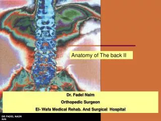
Anatomy of The back II. Examination Of The Back. It is important that: The whole area of the back and legs be examined The shoes be removed. Unequal length of the legs or disease of the hip joints can lead to abnormal curvatures of the vertebral column.

E N D
Examination Of The Back • It is important that: • The whole area of the back and legs be examined • The shoes be removed. • Unequal length of the legs or disease of the hip joints can lead to abnormal curvatures of the vertebral column. • The patient should be asked to walk up and down the examination room so that the normal tilting movement of the pelvis can be observed
As one side of the pelvis is raised • A coronal lumbar convexity develops on the opposite side • A compensatory thoracic convexity on the same side. • When a person assumes the sitting position • The normal lumbar curvature becomes flattened • An increase in the interval between the lumbar spines.
The normal range of movement of the different parts of the vertebral column should be tested. • In the cervical region • Flexion • The patient should be able to touch his or her chest with the chin • About half of the movement is carried out at the atlanto-occipital joints • Extension • The patient should be able to look directly upward • Lateral rotation • The patient should be able to place the chin nearly in line with the shoulder • Half of lateral rotation occurs between the atlas and the axis. • Lateral flexion • In lateral flexion the head can normally be tilted 45° to each shoulder. • It is important that the shoulder is not raised when this movement is being tested.
In the thoracic region • The movements are limited by the presence of the ribs and sternum. • When testing for rotation, make sure that the patient does not rotate the pelvis. • In the lumbar region • Flexion • Extension • Flexion and extension are fairly free • Lateral rotation • Limited by the interlocking of the articular processes • Lateral flexion • Tested by asking the patient to slide, in turn, each hand down the lateral side of the thigh.
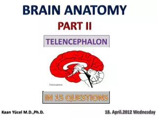
JOINTS OF THE VERTEBRAL COLUMNAtlanto-occipital Joints • Synovial joints • Formed between: • The occipital condyles, on either side of the foramen magnum above • The facets on the superior surfaces of the lateral masses of the atlas below • Movements • Capable of flexion, extension, and lateral flexion; • Do not rotate.
JOINTS OF THE VERTEBRAL COLUMNAtlanto-occipital Joints • Ligaments • Anterior atlanto-occipital membrane: • A continuation of the anterior longitudinal ligament, • Runs as a band down the anterior surface of the vertebral column. • The membrane connects the anterior arch of the atlas to the anterior margin of the foramen magnum . • Posterior atlanto-occipital membrane: • Similar to the ligamentum flavum • Connects the posterior arch of the atlas to the posterior margin of the foramen magnum.
Atlanto-Axial Joints • Three synovial joints; • One is between the odontoid process and the anterior arch of the atlas • The other two are between the lateral masses of the bones. • Movements • There can be extensive rotation of the atlas and thus of the head on the axis.
Atlanto-Axial Joints • Ligaments • Apical ligament: • This median-placed structure connects the apex of the odontoid process to the anterior margin of the foramen magnum. • Alar ligaments: • These lie one on each side of the apical ligament and connect the odontoid process to the medial sides of the occipital condyles. • Cruciate ligament: • Consists of a transverse part and a vertical part. • The transverse part: • Attached on each side to the inner aspect of the lateral mass of the atlas • Binds the odontoid process to the anterior arch of the atlas. • The vertical part: • Runs from the posterior surface of the body of the axis to the anterior margin of the foramen magnum. • Membrana tectoria: • This is an upward continuation of the posterior longitudinal ligament. • It is attached above to the occipital bone just within the foramen magnum. • It covers the posterior surface of the odontoid process and the apical, alar, and cruciate ligaments.
Joints of the Vertebral Column Below the Axis • Joints between Two Vertebral Bodies • The upper and lower surfaces of the bodies of adjacent vertebrae are covered by thin plates of hyaline cartilage. • Sandwiched between the plates of hyaline cartilage is an intervertebral disc of fibrocartilage • The collagen fibers of the disc strongly unite the bodies of the two vertebrae
Joints of the Vertebral Column Below the Axis • Joints between two vertebral arches • Consist of synovial joints between the superior and inferior articular processes of adjacent vertebrae • The articular facets are covered with hyaline cartilage • The joints are surrounded by a capsular ligament.
NERVE SUPPLY OF VERTEBRAL JOINTS • The joints of any particular level receive nerve fibers from two adjacent spinal nerves • The joints between the vertebral bodies are innervated by the small meningeal branches of each spinal nerve • The nerve arises from the spinal nerve as it exits from the intervertebral foramen. • It then reenters the vertebral canal through the intervertebral foramen and supplies • The meninges • Ligaments • Intervertebral discs. • The joints between the articular processes are innervated by branches from the posterior rami of the spinal nerves
DISLOCATIONS OF THE VERTEBRAL COLUMN • Dislocations without fracture occur only in the cervical region • because the inclination of the articular processes of the cervical vertebrae • In the thoracic and lumbar regions, dislocations can occur only if the vertically placed articular processes are fractured. • Dislocations commonly occur between the fourth and fifth or fifth and sixth cervical vertebrae, where mobility is greatest.
DISLOCATIONS OF THE VERTEBRAL COLUMN • In unilateral dislocations the inferior articular process of one vertebra is forced forward over the anterior margin of the superior articular process of the vertebra below. • Because the articular processes normally overlap, they become locked in the dislocated position. • The spinal nerve on the same sideis usually nipped in the intervertebral foramen, producing severe pain. • Fortunately, the large size of the vertebral canal allows the spinal cord to escape damage in most cases.
DISLOCATIONS OF THE VERTEBRAL COLUMN • Bilateral cervical dislocations are almost always associated with severe injury to the spinal cord. • Deathoccursimmediately if the uppercervicalvertebrae are involved because the respiratorymuscles, includingthediaphragm (phrenic nerves C3 to 5), are paralyzed
ANTERIOR AND LATERAL COMPRESSION FRACTURES • Usually caused by an excessive flexion compression type of injury • Take place at • The sites of maximum mobility • The junction of the mobile and fixed regions of the column • Vertebral displacement and spinal cord injury do not occur. • The body of a vertebra in such a fracture is crushed, • The strong posterior longitudinal ligament remains intact • The vertebral arches remain unbroken • The intervertebral ligaments remain intact • When injury causes excessive lateral flexion in addition to excessive flexion, the lateral part of the body is also crushed.
FRACTURE DISLOCATIONS • Usually caused by a combination of a flexion and rotation type of injury • The upper vertebra is excessively flexed and twisted on the lower vertebra • The site is usually where maximum mobility occurs • As in the lumbar region • At the junction of the mobile and fixed region of the column • As in the lower lumbar vertebrae. • Because the articular processes are fractured and the ligaments are torn, the vertebrae involved are unstable, • The spinal cord is usually severely damaged or severed with accompanying paraplegia.
VERTICAL COMPRESSION FRACTURES Jefferson's fracture • In the cervical region, with the neck straight, an excessive vertical force applied from above will cause the ring of the atlas to be disrupted and the lateral masses to be displaced laterally • If the neck is slightly flexed, the lower cervical vertebrae remain in a straight line and the compression load is transmitted to the lower vertebrae, causing disruption of the intervertebral disc and break up of the vertebral body. • Pieces of the vertebral body are commonly forced back into the spinal cord.
VERTICAL COMPRESSION FRACTURES • It is possible for non-traumatic compression fractures to occur in severe cases of osteoporosis and for pathologic fractures to take place. • In the straightened lumbar region, an excessive force from below can cause the vertebral body to break up, with protrusion of fragments posteriorly into the spinal canal.
FRACTURES OF THE ODONTOID PROCESS • Fractures of the odontoid process are relatively common and result from falls or blows on the head • Excessive mobility of the odontoid fragment or rupture of the transverse ligament can result in compression injury to the spinal cord.
FRACTURE OF THE PEDICLES OF THE AXIS(HANGMAN'S FRACTURE) • Severe extension injury of the neck, such as might occur in an automobile accident or a fall, is the usual cause • Sudden overextension of the neck, as produced by the knot of a hangman's rope beneath the chin, is the reason for the common name. • Because the vertebral canal is enlarged by the forward displacement of the vertebral body of the axis, the spinal cord is rarely compressed
SPONDYLOLISTHESIS • The body of a lower lumbar vertebra, usually the fifth, moves forward on the body of the vertebra below • Carries with it the whole of the upper portion of the vertebral column. • The essential defect is in the pedicles of the migrating vertebra. • The pedicles are abnormally formed and accessory centers of ossification are present and fail to unite. • The spine, laminae, and inferior articular processes remain in position
SPONDYLOLISTHESIS • The remainder of the vertebra, having lost the restraining influence of the inferior articular processes, slips forward. • Because the laminae are left behind, the vertebral canal is not narrowed • The nerve roots may be pressed on, causing low backache and sciatica. • In severe cases the trunk becomes shortened, and the lower ribs contact the iliac crest.
Muscles of the Back • Divided into three main groups: • The superficial muscles • Associated with the shoulder girdle • The intermediate muscles • Involved with respiration, • The deep muscles • Belonging to the vertebral column.
The Line Of Gravity • In the standing position it passes through the odontoid process of the axis, behind the centers of the hip joints, and in front of the knee and ankle joints. • when the body is in this position, the greater part of its weight falls in front of the vertebral column. • Therefore the postvertebral muscles of the back are well developed in humans • The postural tone of these muscles is the major factor responsible for the maintenance of the normal curves of the vertebral column.
SUPERFICIAL MUSCLES • The superficial muscles: • The trapezius • Latissimus dorsi • Levator scapulae • Rhomboid minor and major
INTERMEDIATE MUSCLES • The intermediate muscles • The serratus posterior superior • Serratus posterior inferior • Levatores costarum
The deep muscles of the back • The spines and transverse processes of the vertebrae serve as levers that facilitate the muscle actions. • The muscles of longest length lie superficially and run vertically from the sacrum to the rib angles, the transverse processes, and the upper vertebral spines • The muscles of intermediate length run obliquely from the transverse processes to the spines. • The shortest and deepest muscle fibers run between the spines and between the transverse processes of adjacent vertebrae.
The deep muscles of the back may be classified as follows: • Superficial Vertically Running Muscles • Erector spinae • longissimus • Iliocostalis • spinalis
Intermediate Oblique Running Muscles • Semispinalis • Multifidus • Rotators
Deepest Muscles • Interspinales. • Intertransversarii. • Nerve Supply • All the deep muscles of the back are innervated by the posterior rami of the spinal nerves.
SPLENIUS • The splenius is a detached part of the deep muscles of the Back. • It consists of two parts. • The splenius capitis • Arises from • The lower part of the ligamentum nuchae • The upper four thoracic spines • Inserted into • The superior nuchal line of the occipital bone • The mastoid process of the temporal bone. • The splenius cervicis • Similar origin • Inserted into the transverse processes of the upper cervical vertebrae
Deep Fascia of the Back (Thoracolumbar Fascia)The Lumbar Part Of The Deep Fascia • Situated in the interval between the iliac crest and the 12th rib. • It forms a strong aponeurosis • Laterally gives origin to • The middle fibers of the transversus • The upper fibers of the internal oblique muscles of the abdominal wall
Deep Fascia of the Back (Thoracolumbar Fascia)The Lumbar Part Of The Deep Fascia • Medially, the lumbar part of the deep fascia splits into three lamellae. • The posterior lamella: • Covers the deep muscles of the back • Attached to the lumbar spines. • The middle lamella • Passes medially, • Attached to the tips of the transverse processes of the lumbar vertebrae • It lies • In front of the deep muscles of the back • Behind the quadratus lumborum. • The anterior lamella • Passes medially • Attached to the anterior surface of the transverse processes of the lumbar vertebrae; • It lies in front of the quadratus lumborum muscle.
Deep Fascia of the Back (Thoracolumbar Fascia) • In the thoracic region • The deep fascia is attached: • medially to the vertebral spines • laterally to the angles of the ribs. • It covers the posterior surface of the deep muscles of the back. • In the cervical region • The deep fascia is much thinner and of no special importance
Blood Supply of the BackARTERIES • The following arteries supply the structures of the back. • In the cervical region by: • The occipital artery, a branch of the external carotid • The vertebral artery, a branch of the subclavian • The deep cervical artery, a branch of the costocervical trunk, a branch of the subclavian artery • The ascending cervical artery, a branch of the inferior thyroid artery. • In the thoracic region by: • The posterior intercostal arteries • In the lumbar region by: • The subcostal and lumbar arteries. • In the sacral region by • The iliolumbar and lateral sacral arteries • Branches of the internal iliac artery.
VEINS • Complicated plexuses extending along the vertebral column from the skull to the coccyx. • The veins can be divided into • External sinuses within vertebral venous plexus • Lie external to the vertebral column and surround it • Internal vertebral venous plexus • Lie within the vertebral canal • These plexuses freely communicate with the veins in the neck, thorax, abdomen, and pelvis. • Above they communicate through the foramen magnum with the occipital and basilar venous the cranial cavity.
VEINS • The internal vertebral plexus • lies within the vertebral canal but outside the dura mater • It is embedded in areolar tissue • receives tributaries from • the vertebrae by way of the basi-vertebral veins the meninges and spinal cord. • The internal plexus is drained by the inter-vertebral veins • pass outward with the spinal nerves through the inter-vertebral foramina. • joined by tributaries from the external vertebral plexus • drain into the vertebral, intercostal, lumbar, and lateral sacral veins.
VEINS • The external and internal vertebral plexuses: • form a capacious venous network whose walls are thin channels • incompetent valves or are valveless • Free venous blood flow may therefore take place between the skull, the neck, the thorax, the abdomen, the pelvis, and the vertebral plexuses • the direction of flow depending on the pressure differences that exist at any given time between the regions.
VERTEBRAL VENOUS PLEXUS AND CARCINOMA OF THE PROSTATE • Pelvic venous blood enters not only the inferior vena cava but also the vertebral venous plexus and by this route may also enter the skull. • This is especially likely to occur if the intra-abdominal pressure is increased. • The internal vertebral venous plexus is not subject to external pressures when the intra-abdominal pressure rises. • A rise in pressure on the abdominal and pelvic veins would tend to force the blood backward out of the abdominal and pelvic cavities into the veins within the vertebral canal. • The existence of this venous plexus explains how carcinoma of the prostate may metastasize to the vertebral column and the cranial cavity.
Lymph Drainage of the Back • The deep lymph vessels follow the veins • drain into the deep cervical, posterior mediastinal, lateral aortic, and sacral nodes. • The lymph vessels from the skin of the neck drain into the cervical nodes • from the trunk above the iliac crests drain into the axillary nodes • those from below the level of the iliac crests drain into the superficial inguinal nodes
Nerve Supply of the Back • The skin and muscles of the back are supplied in a segmental manner by the posterior rami of the 31 pairs of spinal nerves. • The posterior rami of the 1st , 6th, 7th, and 8th cervical nerves and the 4th and 5th lumbar nerves supply the deep muscles of the back and do not supply the skin. • The posterior ramus of the 2nd cervical nerve (the greater occipital nerve) ascends over the back of the head and supplies the skin of the scalp. • The posterior rami run downward and laterally and supply a band of skin at a lower level than the intervertebral foramen from which they emerge. • Considerable overlap of skin areas supplied occurs so that section of a single nerve causes diminished, but not total, loss of sensation. • Each posterior ramus divides into a medial and a lateral branch