Download
1 / 11
130 likes | 626 Vues
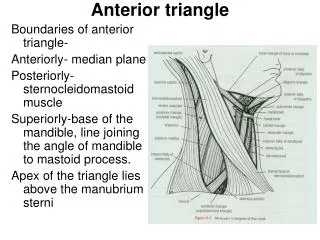
Scalenus Anterior Origin: From the transverse processes of the 3 rd ; 4 th ; 5 th and 6 th cervical vertebrae. Insertion: Into the scalene tubercle on the inner border of the 1 st rib and into the ridge on the upper surface of the 1 st rib.

E N D
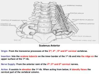
Scalenus Anterior Origin: From the transverse processes of the 3rd; 4th ; 5th and 6th cervical vertebrae. Insertion: Into the scalene tubercle on the inner border of the 1st rib and into the ridge on the upper surface of the 1st rib. Nerve Supply: From the anterior rami of the 4th; 5th and 6th cervical nerves. Action: It assists in elevation the 1st rib. When acting from below, it laterally flexes the cervical part of the vertebral column.
Relation of scalenus anterior muscle Anteriorly: The prevertebral layer of deep cervical fascia, which binds the phrenic nerve down to the anterior surface of the muscle; the superficial cervical and suprascapular arteries which cross the phrenic nerve; and the internal jugular and subclavian veins. Posteriorly: The subcalvian artery; brachial plexus and the cervical dome of the pleura. Medially: The vertebral artery&vein; inferior thyroidartery; thyrocervical trunk; sympathetictrunk and on the left side, thoracic duct. Laterally: The roots of the phrenic nerve unite at the lateral border of the muscle at the level of the cricoid cartilage. Then it descends on its anterior surface. The roots of the brachial plexus & subclavian artery emerge from behind the lateral border of it to enter the posterior triangle of neck.
Scalenus Medius Origin: From the transverse processes of the upper six cervical vertebrae. Insertion: Into the upper surface of the 1st rib behind the groove for the subclavian artery. It lies behind the roots of the brachial plexus and behind the subclavian artery. Nerve Supply: Branches from the anterior rami of the cervical nerves. Action: like scalenus anterior.
Scalenus Posterior Origin: From the transverse processes of the lower cervical vertebrae. Insertion: Into the outer surface of the second rib. Nerve supply: branches from the anterior rami of the lower cervical nerves. Action: Elevates the 2nd rib. When active from below, it laterally flexes the cervical part of the vertebral column.
4 3 2 1 Anterior vertebral ( prevertebral ) muscles 1- Longus coli. 2- Longus capitis. 3- Rectus capitus anterior. 4- Rectus capitus lateralis.
Scaleno-vertebral triangleIts boundaries are theLongus colli ; scalenus anterior and the transverse process of 6th cervical vertebra.It contains 1st part of subclavian artery ; sympathetic chain ; vertebral artery and the thoracic duct ( on left side ).
Cervical plexus It is formed by anterior rami of 1, 2, 3 ; 4 cervical nerves. The rami are joined by connecting branches forming a loops on the levator scapulae and scalenus medius muscles. It is covered by the prevertebral fascia. It is related to the internal jugular vein within the carotid sheath.
C2 • Branches: • 1- Cutaneous • lesser occipital; greater auricular; transverse cutaneous and supraclavicular nerves. • 2- Phrenic nerve to the diaphragm • 2- Muscular • - Prevertebral muscles - Sternocleidomastoid (C2 & C3as proprioceptive ). • - Levator scapule (C3&C4motor • - Trapezius ( C3 & C4 asproprioceptive ). • - A branch of C1 joins the hypoglossal nerve then it leaves it to supply the thyrohoid & geniohyoid. • - Some of C1 fibers (descending branch) unites with the descending cervical nerves of C2& C3 to form the ansa cervicalis which supplies omohyoid; sternohyoid and sternothyroid muscles.
Phrenic Nerve It arises from the 3rd ; 4th and 5th cervical nerves of cervical plexus. It runs vertically downward acrosses the front of the scalenus anterior from its lateral to medial border behind the prevertebral layer of deep cervical fascia. It enters the thorax by passing in front of the subclavian artery and behind the beginning of the brachiocephalic vein.
Relations Anteriorly: The prevertebral layer of the deep fascia; internal jugular vein; superficial cervical and suprascapular arteries; beginning of the brachiocephalic vein and on the left side the thoracic duct. Posteriorly: The scalenus anterior; subclavian artery and the cervical dome of pleura.
Branches: 1- It is the only motor nerve supply to the diaphragm. 2- It contains sympathetic fibers. 3- It contains proprioceptive sensory fibers ( stimulation of the sensory nerve ending in the muscles& tendons and joints ) for the muscles of the diaphragm. 4- It contains sensory fibers for the mediastinal pleura and the pericardium. 5- It contains Sensory fibers for the pleura & peritoneum covering the upper and lower surfaces of the central part of the diaphragm.