Anterior triangle
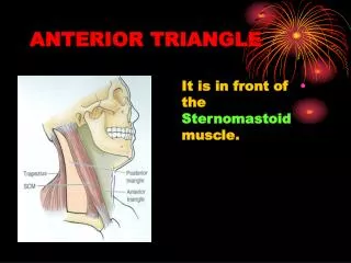
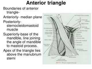
Anterior triangle. Boundaries of anterior triangle- Anteriorly- median plane Posteriorly- sternocleidomastoid muscle Superiorly-base of the mandible, line joining the angle of mandible to mastoid process. Apex of the triangle lies above the manubrium sterni. Anterior triangle. Landmarks

Anterior triangle
E N D
Presentation Transcript
Anterior triangle Boundaries of anterior triangle- Anteriorly- median plane Posteriorly- sternocleidomastoid muscle Superiorly-base of the mandible, line joining the angle of mandible to mastoid process. Apex of the triangle lies above the manubrium sterni
Anterior triangle Landmarks • Mandible • Hyoid bone • Thyroid cartilage • Cricoid cartilage • Trachea
Anterior triangle Skin Free movable Ant cut nerve of C2,C3 Superficial fascia • Platysma • Cervical branch of the facial nerve • Anterior jugular vein • Lymph nodes ( lymph nodes along the AJV, submental nodes lies below the chin & few of them lie along the sternocleidomastoid along the EJV)
Anterior triangle Structures in the anterior median region of the neck Skin- freely movable Superficial fascia- • Upper decussating fibres of the platysma • Anterior jugular vein • Submental lymph nodes • Transverse or anterior cutaneous nerve of neck Deep fascia- Above the hyoid bone its single layer in the median plane but splits on each side to enclose the submandibular salivary gland Between hyoid bone and cricoid cartilage is single layer extending the rt & lt sternoclediomastoid muscles Below the cricoid, fascia splits to enclose the suprasternal space.
Anterior triangle Deep structures lying above the hyoid bone Mylohyoid muscles is overlapped by • Ant belly of digastric muscles • Superficial part of submandibular salivary gland • Mylohyoid nerve & blood vessels • Submental branch of the facial artery • Anterioinferiorly hyoglossus muscles • Intermediate tendon of digastric muscle with its fibrous pully • Bifurcated tendon of the stylohyoid muscle • Hypoglossal nerve • Subhyoid bursa- between the posterior surface of hyoid bone and the thyrohyoid membrane
Anterior triangle Structures lying below the hyoid bone Superficial plane ( infrahyoid muscles)- • Sternohyoid • Sternothyoid • Thyrohyoid • Superior belly of omohyoid Pretracheal fascia- False capsule of the thyroid gland and the suspensory ligament. Inferior thyroid veins lies within the fascia Deep to pretracheal fascia- • Thyrohyoid membrane deep to thyrohyoid muscle & pierced by internal laryngeal nerve and superior thyroid vessels • Thyroid cartilage • Cricothyroid membrane • Arch of the cricoid cartilage • Cricothyroid muscle • Trachea ( covered by isthmus of the thyroid gland from 2to 4th rings) • Carotid sheath • Lt brachiocephalic vein and brachiocephalic artery
Anterior triangle Clinical anatomy- • Enlargement of submental lymph nodes & sublingual dermoid in the submental region • Thyroglaossal cyst and subhyoid bursitis just below the hyoid bone • Goiter, carcinoma of the larynx and enlarged lymph nodes in the suprasternal region • Tracheostomy-retrothyroid region after retracting the isthmus of the thyroid gland. Suprathyroid tracheostomy is liable to stricture & infrathyroid one is difficult due to the depth of the trachea and is also dangerous because numerous blood vessels lie to the trachea here • “Cut throat”- commonly situated just above or below the hyoid bone.
Anterior triangle Subdivision • Triangle is enclosed by suprahyoid muscles- digastric, stylohyoid, mylohyoid & geniohyoid • Infrahyoid muscles- sternohyoid, sternothyroid, thyrohyoid & omohyoid
Anterior triangle Triangle is subdivided by the digastric & superior belly of the omohyoid muscle into • Submental • Digastric • Carotid • Muscular
Anterior triangle Submental triangle- Boundaries- Each side- ant belly of corresponding digastric muscles Base- body of hyoid bone Apex-chin Floor- rt & lt mylohyoid muscles and median raphe uniting them. Contents- • 2-4 submental lymph nodes situated in the superficial fascia between the anterior bellies of the digastric muscles. Drains lymph from superficial tissues below the chin, central part of the lower lip, adjoining gum, anterior part of the floor of the mouth & tip of the tongue • Small submental veins to form anterior jugular vein
Anterior triangle Digastric triangle- Boundaries- Anteroinferiorly- ant belly of digastric Posteroinferiorly- post belly of digastric & stylohyoid Superiorly or base- base of the mandible & line joining the angle of the mandible to the mastoid process Roof- • Skin • Superficial fascia- platysma, cervical branch of the facial nerve & ascending branch of the transverse or anterior cutaneous nerve of the neck • Deep fascia- splits to enclose the submandibular salivary gland Floor- mylohyoid anteriorly& hyoglossus posteriorly, small part of middle constrictor muscle of pharynx appears in the floor Contents- • Structures superficial to mylohyoid- a)superficial part of the submandibular salivay gland, facial vein and submandibular lymph nodes are superficial to it and facial artery is deep to it; b) submental artery & c) mylohyoid nerve and vessels
Anterior triangle 2. Structures superficial to the hyoglossus- submandiblar salivary gland, intermediate tendon of the digastric & stylohyoid and hypoglossal nerve
Anterior triangle Posterior part of the triangle- • Superficial structures are-lower part of the parotid gland & ECA • Deep structures passing between ECA& ICA are- styloglossus, stylopharyngeus, glossopharyngeal nerve,pharyngeal branch of the vagus nerve,styloid process & a part of the parotid gland. • Deepest structures include- ICA, IJV & vagus nerve.
Anterior triangle Carotid triangle Boundaries- Anterosuperiorly- post belly of the digastric muscle & stylohyoid Anteroinferiorly- superior belly of the omohyoid Posteriorly- ant border of the sternocleidomastoid muscle Roof- skin, superficial fascia containing platysma, cervical branch of the facial nerve & transverse cutaneous nerve of the neck Floor- thyrohoid, hyoglossus, & middle and inferior constrictors of the pharynx
Anterior triangle Contents- Arteries- CCA with carotid sinus & carotid body, ICA, ECA with its superior thyroid, lingual, facial, ascending pharyngeal and occipital branches Veins- IJV, CFV draining into the IJV, pharyngeal vein, lingual vein Nerves- vagus nerve, superior laryngeal branch of vagus nerve, dividing into external & internal laryngeal nerves, spinal accessory nerve, hypoglossal nerve & sympathetic chain Carotid sheath and its contents Lymph nodes-deep cervical lymph nodes situated along the IJV and jugulodigastric node below the digastric and jugulo- omohyoid nodes above the inferior belly of the omohyoid.
Anterior triangle CCA- Rt CCA is branch of brachiocephalic artery. Begins in the neck behind the rt sternoclavicular jt Lt CCA is the branch of the Arch of aorta. It begins in the thorax in front of trachea opposite a point little to the left of the center of the manbrium. Ascends to back of the lt sternoclavicular jt & enter the neck. In the neck both have the same course. It runs upwards within the carotid sheath, under cover of the anterior border of the sternocleidomastoid, it lies infront of the lower four cervical transverse processes. At the level of upper border of the thyroid cartilage it ends by dividing into ECA & ICA. Carotid sinus- baroreceptor or pressure receptor and regulates the blood pressure Carotid body-chemoreceptor and responds to the changes in the oxygen & carbondioxide and the pH content of the blood.
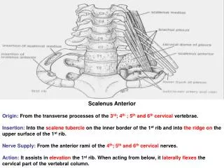
Anterior triangle ECA It lies anterior to the ICA, chief artery which supplies the front of the neck and the face Course & relation- It begins in the carotid triangle at the upper border of the thyroid cartilage opposite the disc betwn the 3rd & 4th cervical vertebra Runs upwards and slightly backwards and laterally & terminates behind the neck of the mandible by dividing into maxillary and superficial temporal arteries. Slightly curved course- it is anteromedial to the ICA in its lower part and anterolateral to ICA in its upper part.
Anterior triangle In carotid triangle- superficial & lies under cover of the sternoclediomastoid. It is crossed superficially by the cervical branch of the facial nerve, hypoglossal nerve & facial, lingual and superior thyroid veins. Deep to it are walls of the pharynx, superior laryngeal nerve which divides into external and internal laryngeal nerves & ascending pharyngeal artery. Above to the triangle- lies deep in the substance of the parotid gland. Within the gland, it is related superficial to the retromandibular vein and facial nerve. Deep to it is ICA and betwn the ECA & ICA are- styloglossus, stylopharyngeus, IX CN, pharyngeal branch of X CN & styloid process and two structures deep to the ICA namely the superior laryngeal nerve & superior cervical sympathetic ganglion.
Anterior triangle Branches Anterior- • Superior thyroid • Lingual • Facial Posterior- • Occipital • Posterior auricular Medial- ascending pharyngeal Terminal- • Maxillary • Superficial temporal