Download
1 / 143
1.43k likes | 1.53k Vues
Learn about boundaries, contents, and important muscles in the neck triangles with Dr. H.A. Jaafar from Al-Nahrain University College of Medicine. Discover the divisions and structures within each triangle for better anatomical understanding.

E N D
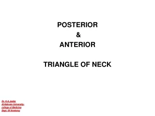
POSTERIOR & ANTERIOR TRIANGLE OF NECK Dr. H.A.Jaafar Al-Nahrain University- college of Medicine Dept. Of Anatomy
Objectives: At the end of this lecture we should be able to know : The boundaries, roof, floor & contents of Anterior triangle The boundaries, contents of posterior triangle The role of Sternomastoid muscle in main anatomical division of the neck. What are the careful and careless area of posterior triangle What are the importance of diagastric and omohyoid muscles as a land marks for subdivision of the two triangular regions of neck.
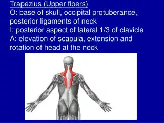
A. Posterior triangle Is bounded by : posterior border of sternocleidomastoid muscle, anterior border of trapezius muscle, superior border of clavicle. roof ………formed by : platysma investing (superficial) layer of deep cervical fascia. floor :…….formed by : splenius capitis levator scapulae muscles anterior, middle, posterior scalene muscles.
Posterior cervical triangle Trapezius Sternocleidomastoid
Is divided into :……. by omohyoid posterior belly occipital triangle. subclavian (supraclavicular) triangle. Contains: accessory nerve, cutaneous branches of cervical plexus, external jugular vein, transverse cervical suprascapular vessels, subclavian vein & artery, Inferior belly of omohyoid, roots and trunks of brachial plexus. nerve to subclavius dorsal scapular, suprascapular, long thoracic nerves.
Triangles of posterior (lateral) region of neck trapezius muscle Sternomastoid muscle
Triangles of posterior (lateral) region of neck clavicle Sternomastoid muscle
Triangles of posterior (lateral) region of neck • Occipital triangle • supraclavicular triangle (greater supraclavicular fossa)
Triangles of posterior (lateral) region of neck • Occipital triangle • supraclavicular triangle (greater supraclavicular fossa)
Sternomastoid muscle trapezius muscle
Sternomastoid muscle trapezius muscle clavicle
Omohyoidmuscle Sternomastoid muscle trapezius muscle clavicle
Omohyoidmuscle Sternomastoid muscle trapezius muscle clavicle Scalenus anterior muscle
Omohyoidmuscle Sternomastoid muscle trapezius muscle Scalenus medius muscle clavicle
Omohyoidmuscle Sternomastoid muscle trapezius muscle Sterno thyroid m clavicle
Omohyoidmuscle Sternomastoid muscle trapezius muscle thyrohyoid m clavicle
Omohyoidmuscle Sternomastoid muscle trapezius muscle Sternohyoid m clavicle
apex trapeziusmuscle Investing layer of deep fascia roof Sternomastoid muscle Middle 1/3 of clavicle – base
Lesser occipital n. External jugular vein Greet auricular n. Transverse nerve of neck Supraclavicular n. Anterior jugular vein Cutaneous nerves and superficial veins
Anterior triangles Suprahyoidmuscles Infrrahyoidmuscles Scalene muscles Prevertebral muscles
B. Anterior triangle Is bounded by: anterior border of sternocleidomastoid, anterior midline of neck, inferior border of mandible. roof :……formed by: platysma investing layer of deep cervical fascia. Is further divided by : omohyoid superior belly digastric anterior and posterior bellies into : digastric (submandibular), submental (suprahyoid), carotid, muscular (inferior carotid) triangles.
Omohyoidmuscle Sternomastoid muscle
Omohyoidmuscle Sternomastoid muscle
Omohyoidmuscle Sternomastoid muscle
Omohyoidmuscle Sternomastoid muscle
apex trapeziusmuscle Investing layer of deep fascia roof Sternomastoid muscle Middle 1/3 of clavicle – base
Anterior triangles Suprahyoidmuscles Infrrahyoidmuscles Prevertebral muscles Scalene muscles
Posterior belly of digastric Stylohyoid Anterior belly of digastric muscle Sternohyoid muscle
Digastric triangle Above …………….lower border of mandible Below & infront …..anterior belly of Digastric muscle Below & behind …..posterior belly of Digastric & stylohyoid muscles . Floor : Anteriorly : ……..mylohyoid muscle Posteriorly ………part of hyoglossusmuscle
Contents of Digastric triangle • submandibular salivary gland • submandibular lymph nodes • lie on the surface of the gland • facial artery • deep to posterior end of submandibular salivary gland • facial vein • lies superficial to submandibular salivary gland • hypoglossalnerve • nerve to mylohyoid muscle
Anterior belly of digastric Digastric triangle • d Posterior belly of digastric Mylohyoid muscle forms the floor of the mouth
C. Hyoid bone……..Is a U-shaped bone consisting of : a median body, lesser horns (cornua) laterally, …………. paired greater horns (cornua) posteriorly……… paired 1. Body Provides for attachments for : Geniohyoidmuscle, mylohyoid, muscle Omohyoidmuscle, sternohyoidmuscle. 2. Greater horn Provides attachments for : middle constrictor, hyoglossus, digastric (anterior and posterior) bellies, stylohyoid, thyrohyoid muscle. 3. Lesser horn Provides attachment for : stylohyoid ligament, which runs from styloid process to lesser horn
D. Styloid process Is a slender projection of variable length extends downward & forward from temporal bone. Gives origin to : three muscles: stylohyoid, styloglossus, Stylopharyngeus two ligaments : stylohyoid stylomandibular.
Carotid triangle boundary : • Behind : …………..sternomastoid muscle • Infront & above : posteriorbelly of digastric muscle • Infront&below : superior belly of omohyoid muscle Floor : infont : hyoglossus muscle ( above ) and the thyrohyoid muscle (below) • Behind: the middle constrictor muscle of the pharynx (above ) and the inferior constrictor muscle of the pharynx (below )
d Posterior belly of digastric Carotid triangle
apex trapeziusmuscle Investing layer of deep fascia roof Sternomastoid muscle Middle 1/3 of clavicle – base
Internal carotid artery External carotid artery Common carotid artery